Periodontitis is a multifactorial, inflammatory disease resulting from an interaction of bacterial infection with immune response. It is an infection of global prevalence, affecting individuals of all ages. Cigarette smoking is an established risk factor for development and progression of periodontitis [1]. According to the “Global Adult Tobacco Survey (GATS) [2] 2010, 34.6% of adults use tobacco, of which cigarette smoking is 5.7%.” The clinical presentation of inflammation is diminished in smokers, such as reduced gingival inflammation due to hyperkeratosis of the gingiva and reduced bleeding due to vaso-constrictive effect of nicotine. Therefore, many a times the periodontal conditions of smokers go unnoticed as tobacco smoking exerts a masking effect on gingival tissues [3,4]. So, early detection and treatment are important to prevent the progression of periodontal disease.
Cigarette smoking results in the disruption of platelet activation and aggregation inducing a great effect on platelet function [5]. When platelets encounter a pathogen, they get activated and release a broad spectrum of immuno-modulatory cytokines, chemokines and other mediators [6]. They modulate the activity of neutrophils resulting in the release of neutrophil extracellular traps, thereby facilitating their recruitment to sites of tissue damage and directly killing the infective cells [7]. Some of the platelet indices like Mean Platelet Volume (MPV) have been found to be biomarkers for platelet activation [8], risk stratification and diagnosis in coronary artery disease patients [9] as it is an inexpensive derivative from complete blood count analysis.
Recently, MPV levels have been studied in various forms of periodontitis and was found to be decreased and the values shifted inversely after active periodontal therapy [10,11]. But there has been no study in smokers with periodontitis. Smokers are more prone for periodontal destruction hence, there is a need to evaluate MPV as an inflammatory marker which would help in early diagnosis and treatment as it is economical with less testing burden for patients as well as the clinicians [12]. In this study, MPV was correlated with salivary C Reactive Protein (CRP), which is one of the established inflammatory markers in periodontitis as well as systemic diseases. So, the primary aim of this study was to assess and correlate MPV and salivary CRP levels among smokers with chronic periodontitis.
Also, to evaluate the clinical periodontal parameters in smokers with or without chronic periodontitis and nonsmokers with or without chronic periodontitis.
Materials and Methods
This was an observational study conducted in the Department of Periodontology, JSS Dental College and Hospital, Mysuru, India, from April 2019-September 2019. The study protocol followed for the present study was approved by the Institutional Review Board Ethical Committee, JSS Dental College and Hospital, Mysore in accordance with the declaration of Helsinki (2013). (JSS/DCH/IEC/MD-07/2017-18).
Study Population: A total of 100 patients were selected by purposive sampling who fulfilled the inclusion and exclusion criteria from the outpatient section of Department of Periodontology (OPD), JSS Dental College and Hospital, Mysuru, India. A written informed consent was taken from the patients before conduction of this study.
Assuming the prevalence of smokers reported to the OPD of periodontology to be 6%, at 95% confidence interval with an error of 0.5, total sample size was computed to be 87. Assuming a 10% non-response, the total sample size was rounded off to 100.
Inclusion criteria: Patients aged 18-50 years with a minimum of 20 teeth were taken into consideration for the study. Smokers were classified as those who have smoked 100 cigarettes in his or her lifetime and nonsmokers were those who had never smoked [13]. Patients with chronic periodontitis, with Pocket Depth (PD) >5mm and attachment loss of at least 4 mm at one or more sites [14] were also recruited in this study.
Exclusion criteria were presence of any other inflammatory oral lesions, former smokers (any adult who had smoked at least 100 cigarettes in his or her lifetime but who had quit smoking at the time of interview) [13], history of periodontal therapy, orthodontic, antimicrobial therapy within the past 6 months, patients on medications known to affect periodontium, any infectious or debilitating diseases.
Clinical Periodontal Parameters
Periodontal examination was done by a single calibrator. Plaque Index (PI) (Silness and loe,1964) [15], Gingival Index (GI) (Loe & Silness, 1963) [15] and Clinical Attachment Levels (CAL) were assessed using William’s periodontal probe while PD was assessed using Florida probe. Four surfaces were evaluated which were “distofacial, facial, mesiofacial, and lingual surfaces.”
Florida probe is a probing and charting software to diagnose periodontal disease, developed by Gibbs CH in 1988 [16]. The greatest advantage is that it has a constant probing pressure of 15 g which is maintained by the coil springs inside the hand piece.
The patients were allotted into four groups:
Group A: Smoker with periodontitis (n=25)
Group B: Smoker without periodontitis (n=25)
Group C: Nonsmoker with periodontitis (n=25)
Group D: Nonsmoker without periodontitis (n=25)
Haematological Analysis for MPV
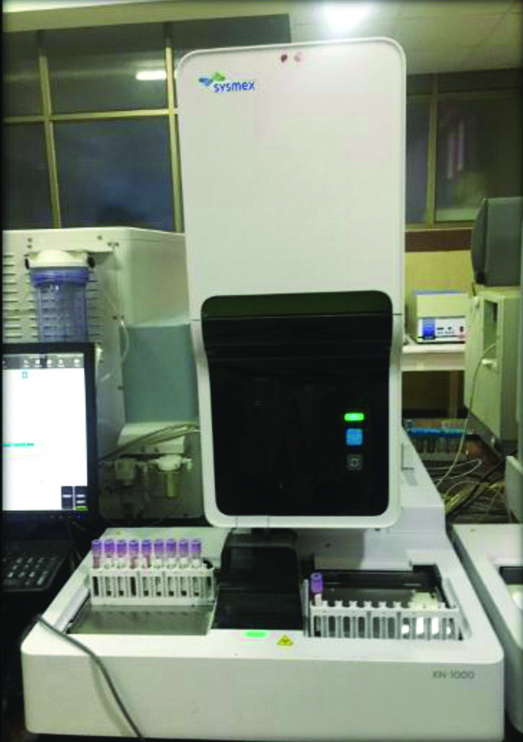
Fasting venous blood sample (2 mL) from the antecubital vein was collected by a trained phlebotomist in Edetate disodium (EDTA), an anticoagulant containing vacutainers. The samples were analysed at Pathology laboratory of JSS Medical college and Hospital within 2-3 hours for complete blood count using an Automated Haematological Analyser (SYSMEX XN-1000) [Table/Fig-1].
Sysmex XN 1000 automated analyser for MPV.
Biochemical Analysis for Salivary CRP
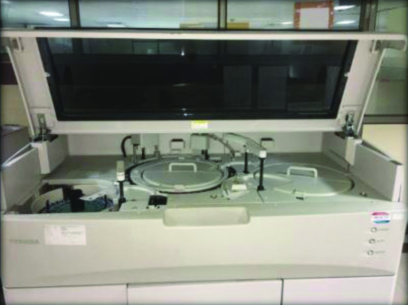
Unstimulated saliva sample (2 mL) was collected in a sterile container and sent to the biochemistry laboratory of JSS Medical College and Hospital for evaluation of salivary CRP levels. The unprocessed samples were aliquoted directly into 1.5 mL Eppendorf tubes and stored at -40°C. All salivary samples were subjected to freeze-thaw cycle to reduce viscosity and to lessen the pipetting errors [17]. All thawed saliva samples were centrifuged (Remi 8-c) at 3000 rpm for 5 minutes to remove cellular debris and to minimise the turbidity of saliva [18]. The supernatant was then transferred into a fresh Eppendorf tube, appropriately labelled and salivary CRP levels were analysed by an automated analyser (TOSHIBA, TBA-120FR) using reagent kits supplied by AGAPPE [Table/Fig-2].
Salivary CRP analysed by TOSHIBA, TBA-120FR.
A questionnaire was handed to the smoker patients (n=50) which included questions related to smoking such as years of smoking, number of cigarettes smoked in a day, chewing any other forms of tobacco, when did they restart smoking after quitting and their willingness to quit smoking. It was done to know the status of smoking and difference in the responses between the groups A and B.
Statistical Analysis
The Statistical package for social science (SPSS version 22) for windows software was used. Quantitative data (PI, GI, PD, CAL, MPV and salivary CRP) were presented as mean and standard deviation while the questionnaire responses were presented as frequencies and percentages. The clinical, haematological and biochemical parameters were observed to be normally distributed. These variables were analysed using one-way ANOVA to test for statistically significant difference between all the groups and Tukey’s post-hoc to test the mean difference in specific groups when compared with each other. Independent sample t-test was done to test for statistically significant difference between the two groups. The correlation between MPV and salivary CRP levels were assessed using Karl Pearson’s product moment correlation co-efficient. Statistical significance was fixed at ≤0.05
Results
The mean age of subjects was found to be 36.80±7.41 years in this study. [Table/Fig-3] depicted that Group A and C consisted of significantly older patients than group B and D (p<0.001), as chronic periodontitis is usually seen in an older population. In [Table/Fig-4], smoking status was investigated from the questionnaire response; majority of the smokers in group A (52%) whereas, in group B (24%) had smoked for >10 years. Smokers in group A (56%) and group B (68%) had smoked 1-10 cigarettes in a day, whereas the remaining 44% in group A and 32% in group B had smoked more than 10 cigarettes in a day. A total of 78% patients were eager to quit smoking, in group A (72%) and group B (84%). None of the participant reported for chewing any form of tabacco. There was no data available on ‘restarting smoking after quitting’. In [Table/Fig-5], the periodontal parameters, haematological and biochemical parameters have been described as mean±SD. Periodontal findings were found to be highest in Group A, followed by Group C, Group B and Group D, respectively. MPV in Group B was found to be highest (11.12±0.72) and least in Group C (9.89±0.53) whereas, Group A depicted highest salivary CRP levels followed by, group B, Group C and Group D, respectively. [Table/Fig-6], depicts a statistically significant (p<0.05) difference in full mouth GI and salivary CRP in all the groups except in PI (p=0.274), PD (p=0.083), CAL (p=0.115) and MPV (p=0.269) between group A and C.
Distribution of age in all the groups (Mean±SD).
| Groups | No. of subjects | Mean age (years) | Standard deviation (SD) |
|---|
| Group A | 25 | 40.70 | 5.35 |
| Group B | 25 | 30.72 | 5.03 |
| Group C | 25 | 41.96 | 6.36 |
| Group D | 25 | 33.76 | 6.32 |
| Total | 100 | 36.80 | 7.41 |
Smoking status (percent) in all the groups.
| Parameters | Group A (n=25) | Group B (n=25) |
|---|
| Smoke years |
| 1-10 years | 48% | 76% |
| >10 years | 52% | 24% |
| No. of cigarettes/day |
| 1-10 | 56% | 68% |
| >10 | 44% | 32% |
| Eagerness to quit |
| Yes | 72% | 84% |
| No | 28% | 16% |
Descriptive statistics of the periodontal, haematological and biochemical parameters (Mean±SD).
| Parameters (Mean±SD) | Group A (n=25) | Group B (n=25) | Group C (n=25) | Group D (n=25) |
|---|
| PI | 2.12±0.35 | 1.67±0.40 | 2.01±0.33 | 1.03±0.34 |
| GI | 1.42±0.15 | 1.22±0.29 | 1.74±0.39 | 0.88±0.24 |
| PD (mm) | 3.85±0.38 | 1.48±0.38 | 3.66±0.38 | 1.82±0.24 |
| CAL (mm) | 2.83±0.39 | - | 2.66±0.46 | - |
| MPV (fL) | 10.06±0.53 | 11.12±0.72 | 9.89±0.53 | 10.44±0.35 |
| Salivary CRP (mg/L) | 7.10±0.62 | 6.07±0.65 | 3.63±0.61 | 0.79±0.36 |
SD: Standard deviation; PI: Plaque index, GI: Gingival index; PD: Probing depth; CAL: Clinical attachment levels; CRP: C-reactive protein; MPV: Mean platelet volume
The t-test for equality of means in between two specific groups and their p-value.
| Groups | “t-test for equality of means” Sig. value |
|---|
| PI | GI | PD | CAL | MPV | CRP |
|---|
| Between A & B | 0.001* | 0.005* | 0.001* | - | 0.001* | 0.001* |
| Between A & C | 0.274 | 0.001* | 0.083 | 0.115 | 0.269 | 0.001* |
| Between A & D | 0.001* | 0.001* | 0.001* | - | 0.005* | 0.001* |
| Between B & C | 0.002* | 0.001* | 0.001* | - | 0.001* | 0.001* |
| Between B & D | 0.001* | 0.001* | 0.001* | - | 0.001* | 0.001* |
| Between C & D | 0.001* | 0.001* | 0.001* | - | 0.001* | 0.001* |
*Statistically significant difference (p<0.05); PI: Plaque index; GI: Gingival index; PD: Probing depth; CAL: Clinical attachment levels; CRP: C-Reactive protein; MPV: Mean platelet volume
[Table/Fig-7] depicts a significant negative relationship between MPV and salivary CRP in group A (r=-0.762, p=0.001), group C (r=-0.973, p=0.001) and group D (r=-0.920, p=0.001), which indicates as MPV level decreases, salivary CRP level linearly increases and a significant positive relationship was observed in smokers without periodontitis, group B (r=0.964, p=0.001) which indicates that as MPV increases, CRP increases linearly and significantly.
Pearson’s product moment correlation between MPV and salivary CRP in individual groups.
| Groups | Variable between | Correlation coefficient (r) | p-value |
|---|
| Group A | MPV & salivary CRP | -0.762 | 0.001* |
| Group B | MPV & salivary CRP | 0.964 | 0.001* |
| Group C | MPV & salivary CRP | -0.973 | 0.001* |
| Group D | MPV & salivary CRP | -0.920 | 0.001* |
*Significant; CRP: C-reactive protein; MPV: Mean platelet volume
Discussion
It is well known that one of the major risk factors for periodontal disease is “cigarette smoking” [1]. However, the nicotine content in the tobacco smoke mask the clinical signs of inflammation, presenting both the patient as well as dental practitioners with an enigma [3,4].
In this study, a statistically significant (p<0.05) difference was noted in full mouth plaque index in all the groups. Highest plaque index was noted in group A (2.12±0.35) followed by group C (2.01±0.33), group B (1.67±0.40) and least in group D (1.03±0.34). But the GI was found to be highest in group C (1.74±0.39) followed by group A (1.42±0.15), group B (1.22±0.29) and least in group D (0.88±0.24). This depicts that despite plaque score being higher in group A, the clinical signs of gingival inflammation were diminished due to the vaso-constrictive effect of nicotine in smokers, which is an agreement with other earlier studies [4,19,20]. Comparing the “pocket depth and CAL in group A and C, both the clinical parameters were observed to be higher in the group A. The difference observed was not statistically significant (p>0.05).
In this preliminary study, the effect of smoking as well as periodontitis was studied on the MPV levels. It was observed that group A and C had significantly lower MPV levels than group B and D. This may be explained due to the migration and consumption of the larger platelets at the inflammation site [21-23]. However, MPV levels when compared in the periodontitis group (A and C) with or without smoking respectively, Group A (10.06±0.53) was noted to be higher than group C (9.89±0.53), but the difference was not statistically significant (p=0.269).
Wang X et al., and Reddy GJ et al., investigated the relationship between MPV and periodontitis and the effect of active periodontal therapy on MPV levels which proved that the MPV levels were decreased in periodontitis patients and the values inversed shift after active periodontal therapy [10,24]. Whereas, in the present study, MPV levels in smokers without periodontitis (Group B) was significantly higher (11.12±0.72), as smoking activates the megakaryocytes to produce larger platelets [25,26].
CRP is one of several proteins that are often referred to as acute phase reactants and is used to monitor changes in inflammation associated with various infectious diseases. It is primarily produced in liver and possesses both pro-inflammatory and anti-inflammatory properties. It is a well known inflammatory marker for chronic periodontitis [27-30].
In present study, it was observed that salivary CRP levels were higher in the smoker group with/without periodontitis (A and B) than nonsmoker group with/without periodontitis (C and D). It was also found that the increased salivary CRP levels were in accordance with the consumption of tobacco smoke. Salivary CRP levels have been assessed as a standard biomarker for periodontitis. Ouellet-Morin I et al., studied to validate a high-sensitivity assay for CRP in saliva as an alternative to serum CRP. He found that salivary CRP allows correct prediction of serum CRP [31].
When salivary CRP levels were compared in periodontitis group (A and C) with or without smoking respectively, CRP in group A was significantly higher (7.10±0.62) than group C (3.63±0.61).
Similar study was done by Shojaee M et al., where salivary CRP levels were assessed in periodontitis and normal subjects using ELISA. He found that salivary CRP concentrations were more among the periodontitis patients indicating a significant relationship between periodontitis and salivary CRP value [32]. Parhar A and Narang S correlated CAL and CRP levels in smokers and nonsmokers with chronic generalised periodontitis and found that CRP was significantly increased in smokers with chronic periodontitis and a strong positive significant relationship was observed between CAL and CRP [33].
Assessment of salivary biochemical parameters among cigarette smokers with chronic periodontitis done by Gazy Y et al., found that salivary CRP was higher in the smokers when compared to nonsmokers [34]. Azar R and Richard A did a pilot study on salivary CRP levels in the context of Tobacco Smoke Exposure (TSE) in healthy youth with an aim to validate salivary CRP against serum CRP. Salivary CRP was measured using ELISA method and was found to be elevated and associated with active smoking in healthy youth. They evidenced the existence of a dose-response relationship between the TSE status and salivary CRP levels [35]. Raval RD et al., evaluated and compared the periodontal disease and smoking as a parallel risk factor for systemic health by gauging the serum CRP levels and evidenced that highest level of CRP was found in smokers with periodontitis followed by non-smokers with periodontitis and smokers without periodontitis. Former smokers had minimum CRP compared to the other groups [36].
A significant negative correlation was observed between MPV and salivary CRP in group A (r=0.762, p=0.001), C (r=0.973, p=0.001) and D (r=0.920, p=0.001), which indicates that as MPV decreases, salivary CRP increases. But, a positive correlation was observed in group B (r=0.964, p=0.001) which indicates that as MPV increases, CRP increases linearly and significantly. The use of MPV as a diagnostic tool among smokers with chronic periodontitis remains uncertain and there is lack of literature to support this result.
From the questionnaire response, a total of 78% patients were eager to quit smoking. Therefore, tobacco cessation therapy should be emphasised by the dental practitioners.
Limitation(s)
Since this was an observational study, the efficacy of MPV as an inflammatory marker based on treatment outcomes could not be evaluated. Platelets collected into EDTA anticoagulants undergo time dependent platelet swelling and activation [37].
Conclusion(s)
In this preliminary study, it was observed that MPV levels are significantly decreased in smokers and nonsmokers with periodontitis. A significant negative correlation was found between MPV and salivary CRP levels in smokers with chronic periodontitis. Nevertheless, the use of MPV as a diagnostic tool among smokers with chronic periodontitis remains uncertain and further high quality epidemiological studies are much needed.
There has been a paucity of literature evaluating salivary CRP using fully automated analyser. Thus, future studies must be carried out with larger sample size to standardise the value.
SD: Standard deviation; PI: Plaque index, GI: Gingival index; PD: Probing depth; CAL: Clinical attachment levels; CRP: C-reactive protein; MPV: Mean platelet volume
*Statistically significant difference (p<0.05); PI: Plaque index; GI: Gingival index; PD: Probing depth; CAL: Clinical attachment levels; CRP: C-Reactive protein; MPV: Mean platelet volume
*Significant; CRP: C-reactive protein; MPV: Mean platelet volume