Hypohidrotic Ectodermal Dysplasia
Chiranjit Ghosh1, Shabnam Zahir2, Shyamal Bar3
1 Clinical Tutor, Department of Paediatric and Preventive Dentistry, North Bengal Dental College and Hospital, Kolkata, West Bengal, India.
2 Professor and Head, Department of Paediatric and Preventive Dentistry, Guru Nanak Institute of Dental Sciences and Research, Kolkata, West Bengal, India.
3 Professor, Department of Orthodontics, Burdwan Dental College and Hospital, Burdwan, West Bengal, India.
NAME, ADDRESS, E-MAIL ID OF THE CORRESPONDING AUTHOR: Dr. Shabnam Zahir, 15 Kabi Tirtha Sarani, Ripples 6 E, Kolkata, West Bengal, India.
E-mail: dr.shabnam.zahir@gmail.com
Anodontia, Hypohidrosis, Hypotrichosis, Oral rehabilitation
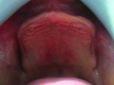
A six-year-old male reported to the Paediatric Dental Department with the complaint of missing teeth from birth, inability to eat and problem in speech. He was a student of grade one with normal intelligence. There was no history of similar illness in his parents and his elder sibling. He gave history of absence of sweating even in summer and frequent rise of body temperature. The patient presented with thin built, dry, wrinkled skin, fine, sparse, light coloured scalp hair, scanty eyebrow and eyelashes, frontal bossing, depressed nasal bridge, full and everted lips, hyper pigmented periocular area [Table/Fig-1]. Finger and toe nails appeared normal [Table/Fig-2,3]. Intraoral clinical examination revealed absence of any deciduous dentition and upper and lower edentulous knife edge ridges deficient in height and width [Table/Fig-4,5]. Salivation and lacrimation was normal. Absence of any developing tooth bud of permanent dentition was confirmed by orthopantomogram [Table/Fig-6]. Based on above clinicoradiological findings a diagnosis of Hypohidrotic Ectodermal Dysplasia (HED) was made.
Extraoral feature is classical old man facies including sparse, fine blond hair with abnormal texture of the scalp, sparse eyebrows and eyelashes, prominent forehead, depressed nasal bridge, midface hypoplasia, protuberant lips, wrinkled and hyperpigmented periocular skin.

Toe nails with no defects.

Finger nails with no obvious defect.

Intraoral feature include complete anodontia of upper arch with hypoplastic alveolar bone with knife edge morphology.

Intraoral feature is complete anodontia of lower arch with hypoplastic alveolar bone with knife edge morphology.

Orthopantomogram reveals absence of any developing tooth bud of permanent dentition.

A removable temporary complete denture were planned keeping in mind the requirement of aesthetics, mastication, speech and overall psychological development, which will be modified by relining or replacing in regular interval as per need of skeletal growth of the child.
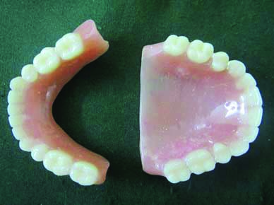
Primary impression was made by impression compound employing the smallest stock tray. Special tray was fabricated and final impression was taken by rubber base impression material. On the master cast occlusal rim were fabricated with wax and appropriate vertical dimension and jaw relation was recorded. Due to unavailability of paediatric mould teeth, smallest size of teeth was chosen to simulate the natural dentition of a six-year-old child. After wax trial the dentures were fabricated in heat cured acrylic resin [Table/Fig-7]. Post-insertion instruction like denture removal at night, storage in water and follow-up after every six months for adjustment or replacement, was given to the patient and parent. Retention of maxillary denture was good but in case of mandible it was compromised and created a problem for the patient. So denture adhesive (Fixodent) was prescribed for mandibular denture. Patient’s aesthetics improved remarkably, he was very happy and was followed-up for one year [Table/Fig-8]. Patient was further referred to Paediatrics Department of a Medical College for speech therapy and other necessary medical management of the case.
Fabricated removable acrylic complete denture for upper and lower dental arch.

Patient wearing removable acrylic complete denture for upper and lower dental arch.

HED/(Christ-Siemens-Touraine syndrome) one of the most common variety of Ectodermal Dysplasia (ED) is a rare genetically determined ectodermal disease characterised by classic triad of hypotrichosis, anodontia/oligodontia, and hypohidrosis/anhidrosis. HED is generally inherited in X-linked recessive manner with classical clinical features expressed in males. Females have few to no symptoms. The prevalence of HED is found to be 1:10,000 and 1:100,000 male live births [1]. It was first described by Thurman in 1848 [1] and the term hereditary ectodermal dysplasia was introduced by Weech (1929) [1]. He also suggested the term anhidrotic (inability to perspire). Felsher (1944) modified it to hypohidrotic as the person with hypohidrotic did not show complete absence but only decreased sweat glands [1].
HED is most commonly diagnosed by delay in eruption of teeth as it happened in our case. Clinical features of HED include thin, scanty hair on the scalp, eyebrows and eyelashes, skin appearing dry and wrinkled, prominent forehead but depressed nasal bridge and protuberant dry and cracked lips, defective toe and finger nail, complete or partial anodontia [2]. Affected individuals typically display heat intolerance because of a reduced number of sweat glands. The periocular skin surrounding the eye may appear wrinkled with hyper pigmentation. Midface hypoplasia with protuberant lips is very common features in patient with HED. Most of these features were seen in the present case with easily recognisable classical facies, also referred to as an old man facies. Skin biopsy and karyotyping would have given a confirmatory diagnostic report, which could not be performed in the present case because of poor socio-economic status of the patient. Alopecia Areata, Rothmund-Thomson syndrome, Focal Dermal Hypoplasia syndrome, Aplasia Cutis Congenita, Pachyonychia Congenita and Werner’s syndrome, Incontinentia Pigmenti, Naegeli-Franceschetti-Jadassohn syndrome are some of the differential diagnosis of HED [3].
Author Declaration:
Financial or Other Competing Interests: None
Was informed consent obtained from the subjects involved in the study? Yes
For any images presented appropriate consent has been obtained from the subjects. Yes
Plagiarism Checking Methods: [Jain H et al.]
Plagiarism X-checker: May 24, 2021
Manual Googling: Aug 17, 2021
iThenticate Software: Sep 10, 2021 (8%)
[1]. Deshmukh S, Prashanth S, Ectodermal dysplasia: A genetic reviewInt J Clin Pediatr Dent 2012 5(3):197-202.10.5005/jp-journals-10005-116525206167 [Google Scholar] [CrossRef] [PubMed]
[2]. Sen S, Das S, Sharma S, Complete phenotypic expression of hypohidrotic ectodermal dysplasia in a female patientIndian J Dermatol 2020 65:540-41.10.4103/ijd.IJD_728_1833487719 [Google Scholar] [CrossRef] [PubMed]
[3]. Singh GP, Saxena V, Hypohidrotic ectodermal dysplasiaMed J Armed Forces India 2015 71(Suppl2):S530-33.10.1016/j.mjafi.2014.05.00626843773 [Google Scholar] [CrossRef] [PubMed]