Villonodular Synovitis of the Index Finger: A Case Report
Anteshwar Birajdar1, Talha Muneer Muneer Mohammed2, Sahil Chowdhary3, Raj Kamble4
1 Associate Professor, Department of Orthopaedics, Dr. D. Y. Patil Medical College, Pune, Maharashtra, India.
2 Resident, Department of Orthopaedics, Dr. D. Y. Patil Medical College, Pune, Maharashtra, India.
3 Resident, Department of Orthopaedics, Dr. D. Y. Patil Medical College, Pune, Maharashtra, India.
4 Resident, Department of Orthopaedics, Dr. D. Y. Patil Medical College, Pune, Maharashtra, India.
NAME, ADDRESS, E-MAIL ID OF THE CORRESPONDING AUTHOR: Dr. Talha Muneer Muneer Mohammed, Resident, Department of Orthopaedics, Dr. D. Y. Patil Medical College, Pune-411018, Maharashtra, India.
E-mail: talha.m1432@gmail.com
Villonodular Synovitis (VNS) is a rare condition characterised by abnormal growth of synovial tissue and accumulation of haemosiderin within joints, tendon sheaths and bursae. While the exact aetiology remains unclear, chronic inflammation is a leading theory. Although VNS typically affects large joints, the present case report highlights the possibility of its occurrence in smaller joints like the index finger. Early diagnosis and surgical intervention are crucial for effective management of VNS. The present case report underscores the importance of considering VNS in the differential diagnosis of soft-tissue masses, even in atypical locations. The present case report describes a 24-year-old male presenting with a decade-long history of pain and swelling in his right index finger. Despite initial radiographic findings being normal, Magnetic Resonance Imaging (MRI) revealed a well-defined soft-tissue mass involving the proximal and middle phalanx, flexor tendons and annular pulley. The presence of haemosiderin deposition within the mass was evident on MRI. The patient underwent open synovectomy, during which a brownish nodular synovium was observed, further supporting the diagnosis of VNS. Histopathological Examination (HPE) of the removed synovial tissue confirmed the presence of haemosiderin granules and haemosiderin-laden macrophages, solidifying the diagnosis of VNS. Post-surgery, the patient initiated a regimen of passive flexion and extension exercises for 15 days. At the 12-month follow-up, there were no clinical or radiological signs of recurrence and the patient had regained full range of motion.
Rare disease, Surgical treatment, Synovectomy, Synovium
Case Report
A 24-year-old male presented with a decade-long history of pain and oedema in his right index finger. The swelling progressively enlarged over time, accompanied by challenges in flexing and extending the finger. The swelling exhibited a diffuse and lobulated consistency, measuring 35×18×23 mm [Table/Fig-1]. The patient had no history of any co-morbidities.
The skin above showed no signs of inflammation. There was no involvement of any other joints.
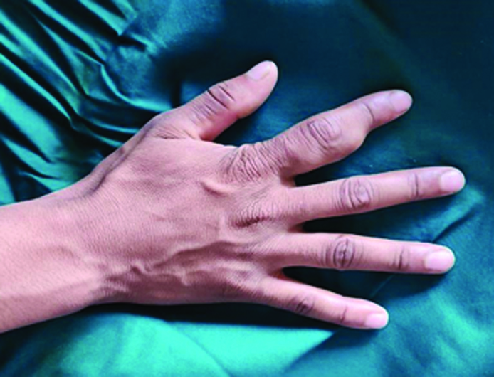
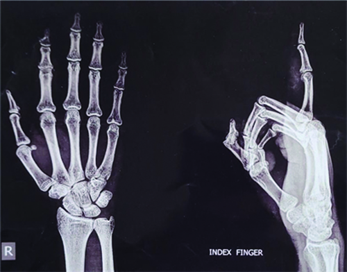
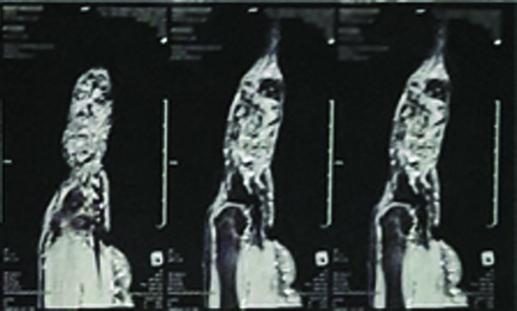
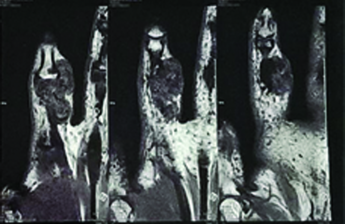
A radiograph of the right index finger revealed no abnormalities in the bone structure [Table/Fig-2]. MRI showed a well-defined lobulated mass of soft tissue observed on the volar aspect of the right index finger, spanning the area near the proximal and middle phalanx as well as the Proximal Interphalangeal (PIP) joint [Table/Fig-3]. The mass also involved the flexor digitorum superficialis and profundus tendons, along with the third annular pulley. Additionally, it surrounded the volar plate of the PIP joint. The lesion extended proximally, spreading between the flexor tendon and the head and body of the proximal phalanx [Table/Fig-4].
Radiographs of the right index finger were taken, revealing no abnormalities in the bone structure.
MRI of right index finger showing a lobulated soft-tissue mass on the volar aspect of the right index finger, extending across the proximal and middle phalanx and the Proximal Interphalangeal (PIP) joint.
MRI of the right index finger showing the mass involving the flexor digitorum superficialis and profundus tendons and the third annular pulley. Evidence of blooming can be seen on the GRE images.
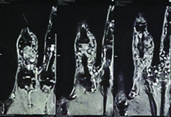
Evidence of blooming on Gradient Recalled Echo (GRE) images suggested the presence of blood products within the lesion [Table/Fig-5]. No signs of cortical irregularities or marrow signal abnormalities were detected in the underlying bones. Furthermore, there was no significant surrounding inflammation noted, such as oedema or infiltration of the soft tissue. All these characteristics aligned with the diagnosis of pigmented VNS.
MRI of the right index finger showing the mass with evidence of blooming can be seen on the GRE images.
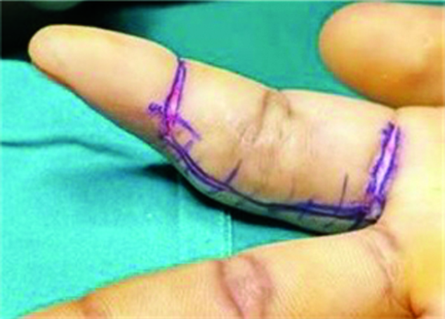
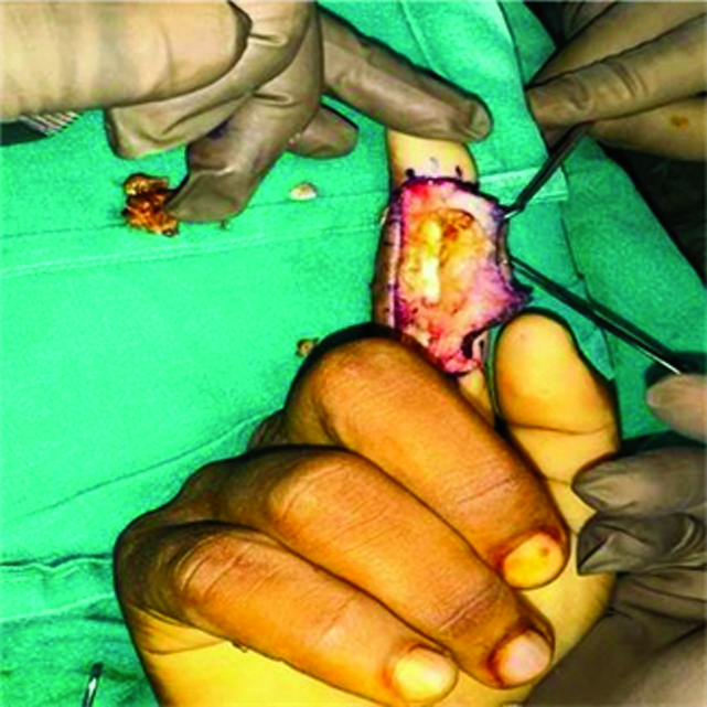
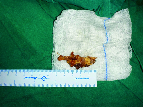
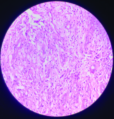
Following the patient‖s written informed consent, he underwent surgery. A total synovectomy was performed via a 6 cm medial incision on the index finger [Table/Fig-6]. Intraoperatively, a brownish nodular synovium was observed, further indicating the presence of VNS [Table/Fig-7]. The synovial tissue was excised during the procedure. The removed synovial tissue [Table/Fig-8] underwent histopathological examination, revealing haemosiderin granules and haemosiderin-laden macrophages scattered throughout the tissue as depicted in the histopathological slides [Table/Fig-9]. These findings collectively confirmed the diagnosis of VNS as reported by the pathologist.
Demonstration of the incision taken.
Intraoperative image of the brown nodular synovium.
Postoperative image of the excised synovium.
Microscopic examination revealing haemosiderin granules and haemosiderin-laden macrophages scattered throughout the tissue. (H&E stain, 100x).
After the surgery, the patient began a regimen of passive flexion and extension exercises for 15 days. Subsequent follow-up appointments were scheduled and at the 12-month evaluation, there were no clinical or radiological signs of recurrence. Additionally, the patient had regained full range of motion.
Discussion
The VNS is characterised by abnormal growth of synovial tissue and accumulation of haemosiderin within joints, tendon sheaths and bursae. The exact aetiology of VNS remains uncertain, with proposed theories including inflammation, neoplastic processes and trauma-induced haemorrhage. This condition predominantly affects large joints and is known for its potential for local aggressiveness and recurrence. The knee is the most frequently affected joint, followed by the hip and ankle. However, VNS can also infrequently involve small joints [1]. Herein, the authors report a unique instance of VNS occurring in the index finger, which was effectively managed through complete tenosynovectomy, tumour resection and tendon reinsertion. Subsequent to the intervention, the patient had full range of motion and there were no signs of recurrence at the 12-month follow-up.
The VNS is a benign neoplastic process affecting the synovium among patients in their third to fifth decades of life. It predominantly presents with intra-articular involvement, with an estimated yearly incidence of 1.8 patients per million [2]. It can manifest in two forms: localised and diffuse. The localised type is distinguished by focal synovial involvement, with either nodular or pedunculated masses, whereas the diffuse form affects the whole synovium. Diffuse intra-articular VNS can be locally aggressive, involving muscles, tendons, bone and skin. When compared to the localised type, diffuse VNS has a higher recurrence risk and is more difficult to treat [3]. The World Health Organisation (WHO) defines diffuse VNS with complete intra-articular involvement under the category of diffuse-type giant cell tumours [4].
The aetiology of VNS remains unclear. While disrupted lipid metabolism is considered a potential mechanism, current research predominantly supports chronic inflammation as the primary process responsible for developing VNS [5-7]. Pain and oedema are the most common clinical signs associated with intra-articular VNS, with joint dysfunction and soft-tissue mass being less common. The duration of these symptoms can be variable [8,9].
The present case was particularly difficult to diagnose due to the uncommon location of the swelling. Clinically, physicians can confuse such masses with common pathologies like tendon tears or tenosynovitis, thus necessitating the use of radiological and pathological modalities [10]. Radiographic imaging can be normal or indicate effusion within the joint and soft-tissue masses that seem hyperdense if the haemosiderin deposition is significant. The presence of bone erosion on X-ray films can be characteristic of VNS. MRI is the preferred diagnostic modality for the disease.
The VNS Magnetic Resonance scans exhibit varied signal intensity according to the percentage of fluid, lipids, haemosiderin and cellular components. Haemosiderin, due to its paramagnetic behaviour, exhibits deep hypointensities on both T1- and T2-weighted images [11]. On histopathologic analysis, VNS lesions consist of villi containing vascular channels that are closely packed with multinucleated giant cells and macrophages containing haemosiderin and lipids [12]. The best surgical option for localised VNS is marginal excision. However, diffuse VNS requires near-complete or entire synovectomy for effective treatment. Arthroscopic synovectomy may have numerous benefits over the open strategy, but it is linked with a higher probability of recurrence in diffuse VNS. External beam radiation offers a high possibility of managing partially resected and unresectable VNS. Additional treatment alternatives include instillation of radioactive isotopes within the joint cavity or cryosurgical surface spray [12].
In the current case, the patient was a young male showing no involvement of hip and knee. In a study by Xie GP et al., 237 cases found that VNS shows a female predominance, occurs mostly between 20-40 years and favours the knee and hip, recurrence is frequent, particularly in the knee and serum Erythrocyte Sedimentation Rate (ESR) and C-reactive Protein (CRP) may be elevated in some patients [13].
The present patient only showed involvement of one joint and no spread or recurrence. In a study by McKean D et al., they report a rare case of polyarticular extension of VNS to contiguous joints via villonodular tenosynovitis, which was not observed in the present study [14].
The present case was diagnosed only after HPE of the excised sample and would have benefited from prior to cytological examination. A case report by Bhatnagar K et al., presented a young female with VNS involving the thumb was treated by in toto excision and the Fine Needle Aspiration Cytology (FNAC) performed prior to surgery matched with the histopathological findings confirmed from the excised sample [15].
Conclusion(s)
The authors present a rare case of VNS occurring in the index finger, successfully managed with open synovectomy. Despite its unusual location, complete excision led to a favourable outcome with no signs of recurrence at the 12-month follow-up. This highlights the importance of considering VNS in the differential diagnosis of soft-tissue masses, even in atypical locations and underscores the efficacy of surgical intervention in achieving long-term symptom resolution and functional recovery.
Author Declaration:
Financial or Other Competing Interests: None
Was informed consent obtained from the subjects involved in the study? Yes
For any images presented appropriate consent has been obtained from the subjects. Yes
Plagiarism Checking Methods: [Jain H et al.]
Plagiarism X-checker: Jul 30, 2024
Manual Googling: Oct 19, 2024
iThenticate Software: Oct 21, 2024 (9%)
[1]. Jaffe HL, Lichtenstein L, Sutro CJ, Pigmented villonodular synovitis, bursitis and tenosynovitisArch Pathol 1941 31:731-65. [Google Scholar]
[2]. Murphey MD, Rhee JH, Lewis RB, Fanburg-Smith JC, Flemming DJ, Walker EA, Pigmented villonodular synovitis: Radiologic-pathologic correlationRadiographics 2008 28(5):1493-518.10.1148/rg.28508513418794322 [Google Scholar] [CrossRef] [PubMed]
[3]. Tyler WK, Vidal AF, Williams RJ, Healey JH, Pigmented villonodular synovitisJ Am Acad Orthop Surg 2006 14(6):376-85.10.5435/00124635-200606000-0000716757677 [Google Scholar] [CrossRef] [PubMed]
[4]. Hong CM, Hing LT, Acute knee pain in a child due to pigmented villonodular synovitisJ Orthop Case Rep 2015 5:78-80. [Google Scholar]
[5]. Granowitz SP, D‖Antonio J, Mankin HL, The pathogenesis and long-term end results of pigmented villonodular synovitisClin Orthop Relat Res 1976 (114):335-51.10.1097/00003086-197601000-00042 [Google Scholar] [CrossRef]
[6]. Sakkers RJ, de Jong D, van der Heul RO, X-chromosome inactivation in patients who have pigmented villonodular synovitisJ Bone Joint Surg Am 1991 73(10):1532-36.10.2106/00004623-199173100-000111748702 [Google Scholar] [CrossRef] [PubMed]
[7]. Flandry F, Norwood LA, Pigmented villonodular synovitis of the shoulderOrthopedics 1989 12(5):715-18.10.3928/0147-7447-19890501-102657682 [Google Scholar] [CrossRef] [PubMed]
[8]. Kransdorf MJ, Murphey MD, Synovial tumorsIn: Imaging of Soft-Tissue Tumors 2006 Philadelphia, PALippincott Williams and Wilkins:381-436. [Google Scholar]
[9]. Myers BW, Masi AT, Pigmented villonodular synovitis and tenosynovitis: A clinical epidemiologic study of 166 cases and literature reviewMedicine (Baltimore) 1980 59(3):223-38.10.1097/00005792-198005000-00004 [Google Scholar] [CrossRef]
[10]. Kinra P, Varghese B, Pigmented villonodular synovitis: Dorsum footMed J Armed Forces India 2007 63(3):294-96.10.1016/S0377-1237(07)80163-827408025 [Google Scholar] [CrossRef] [PubMed]
[11]. Kim IK, Cho HY, Cho HW, Seo JH, Lee DH, Peng W, Pigmented villonodular synovitis of the temporomandibular joint - computed tomography and magnetic resonance findings: A case reportJ Korean Assoc Oral Maxillofac Surg 2014 40(3):140-46.10.5125/jkaoms.2014.40.3.14025045642 [Google Scholar] [CrossRef] [PubMed]
[12]. Mendenhall WM, Mendenhall CM, Reith JD, Scarborough MT, Gibbs CP, Mendenhall NP, Pigmented villonodular synovitisAm J Clin Oncol 2006 29(6):548-50.10.1097/01.coc.0000239142.48188.f617148989 [Google Scholar] [CrossRef] [PubMed]
[13]. Xie GP, Jiang N, Liang CX, Zeng JC, Chen ZY, Xu Q, Pigmented villonodular synovitis: A retrospective multicenter study of 237 casesPLoS One 2015 10(3):e0121451PMCID: PMC437055810.1371/journal.pone.012145125799575 [Google Scholar] [CrossRef] [PubMed]
[14]. McKean D, Thomee E, Papanikitas J, Lee LYW, Yoong P, Yanny S, Polyarticular extension of pigmented villonodular synovitis to contiguous joints via pigmented villonodular tenosynovitisBJR Case Rep 2016 2(2):20150404PMCID: PMC618089910.1259/bjrcr.2015040430363581 [Google Scholar] [CrossRef] [PubMed]
[15]. Bhatnagar K, Bawa AS, Narang V, Kaur P, Pigmented villonodular synovitis of thumb-A cytological diagnosisJ Clin Diagn Res 2017 11(6):ED18-ED20.Epub 2017 Jun 1. PMCID: PMC553537510.7860/JCDR/2017/28184.1009928764182 [Google Scholar] [CrossRef] [PubMed]