Unfused Crossed Renal Ectopia with Ipsilateral Anorchia–A Rare Entity
Bijit Lodh1, Vijayendra Kanwar2, A.K. Kaku Singh3, S. Rajendra Singh4
1 M.Ch. Trainee, Department of Urology, Regional Institute of Medical Sciences (RIMS), Lamphelpat, Imphal, Manipur-795004, India.
2 M.Ch. Trainee, Department of Urology, Regional Institute of Medical Sciences (RIMS), Lamphelpat, Imphal, Manipur-795004, India.
3 Associate Professor, Department of Urology, Regional Institute of Medical Sciences (RIMS), Lamphelpat, Imphal, Manipur-795004, India.
4 Professor & Head, Department of Urology, Regional Institute of Medical Sciences (RIMS), Lamphelpat, Imphal, Manipur-795004, India.
NAME, ADDRESS, E-MAIL ID OF THE CORESPONDING AUTHOR: Dr. Bijit Lodh, M.Ch. Trainee, Department of Urology, Regional Institute of Medical Sciences (RIMS), Lamphelpat, Imphal, Manipur-795004, India.
Phone: 8974504215,
E-mail: drblodh@yahoo.co.in
The reported incidence of unfused crossed renal ectopia is 1 in 75,000 autopsies. A majority of such cases go unnoticed, as they remain asymptomatic and are found incidentally at autopsy. We have presented such a case for its rarity, unusual presentation and association, and have discussed it in light of the available literature. In this report, a 7–year–old boy was pre–operatively diagnosed as having left crossed renal ectopia with a cryptorchid left testis. At surgery, we noticed an ectopic, hydronephrotic left kidney with an absent left testis. Left Nephrectomy was performed, based on findings of a DTPA scan which was done. Post–operative course was uneventful. Histopathology revealed chronic pyelonephritis without any evidence of dysplastic changes.
Crossed renal ectopia, DTPA, Pyelonephritis
Introduction
A crossed renal ectopia is one of the rarest, but second most common congenital fusion anomaly of kidney. Ninety percent of crossed ectopic kidneys are fused to their ipsilateral mates, whereas unfused ectopias constitute only 10 % of such cases. This anomaly is more common in males, with a ratio of 2:1. Left-to-right ectopia is seen three times more frequently than the opposite [1]. Associated genital anomalies are very rarely seen in patients with unfused crossed renal ectopias. Here, we have reported a case of an unfused crossed renal ectopia for its rare occurrence and unusual coexistence with anorchia.
Case Report
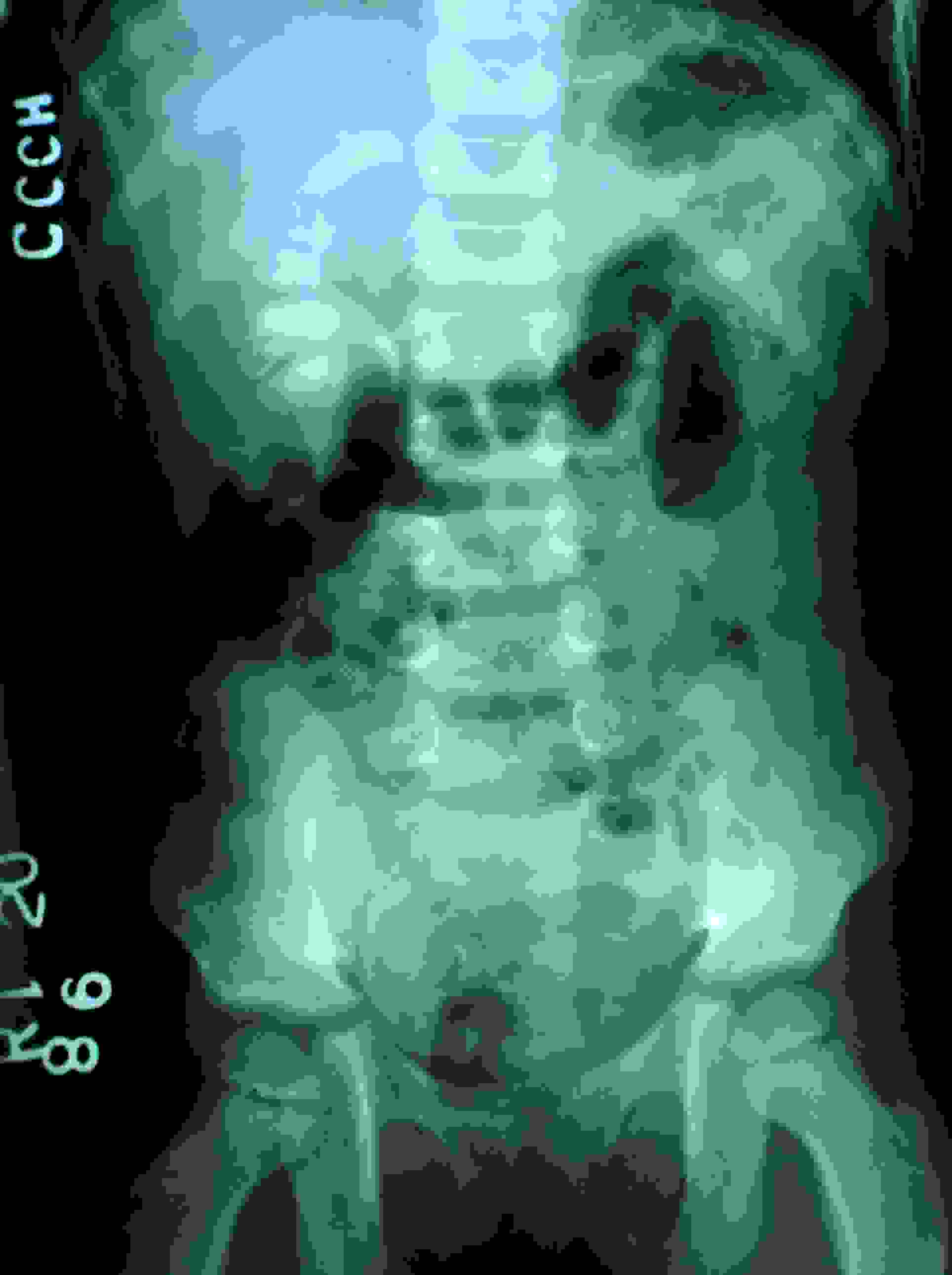
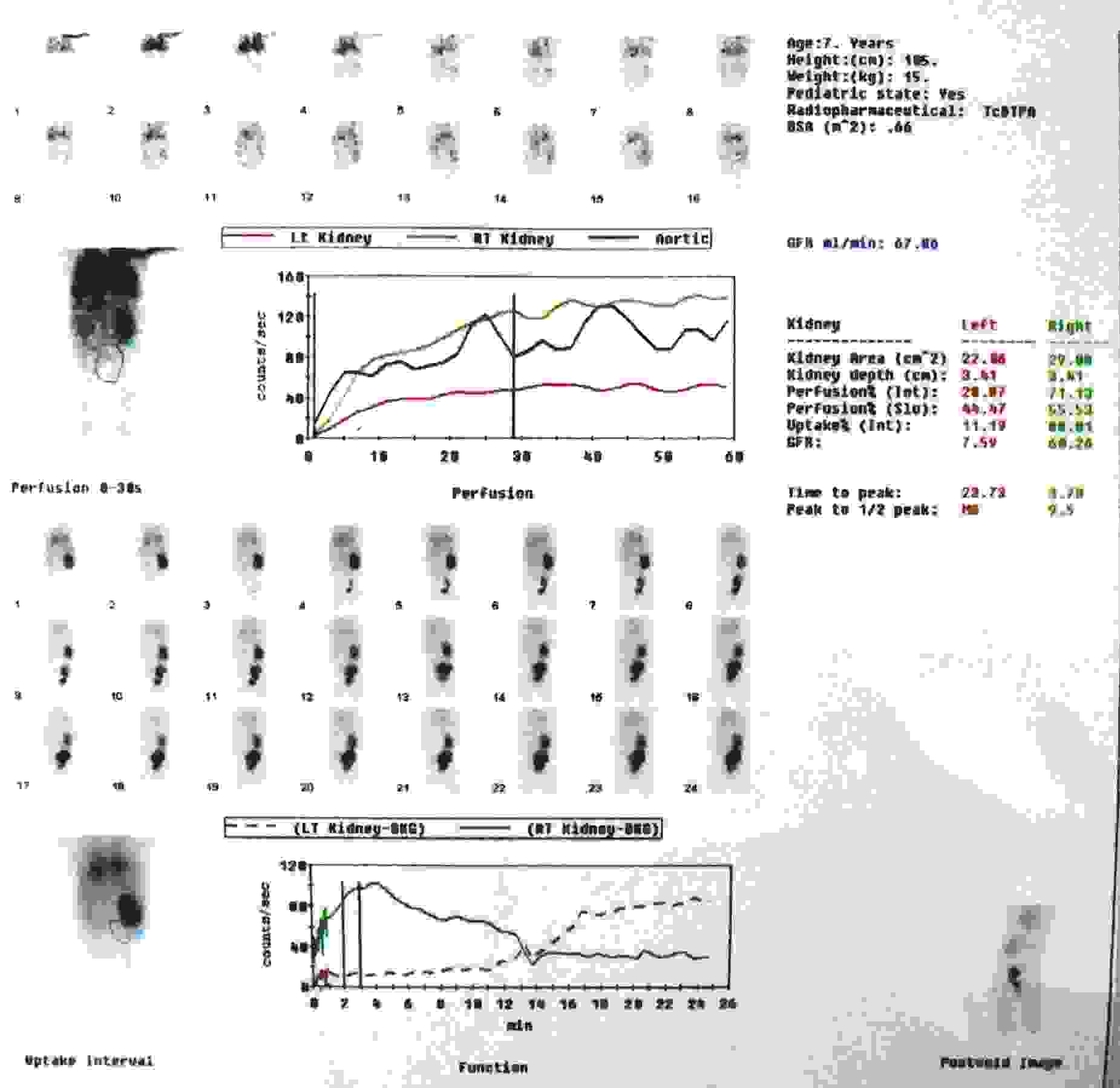
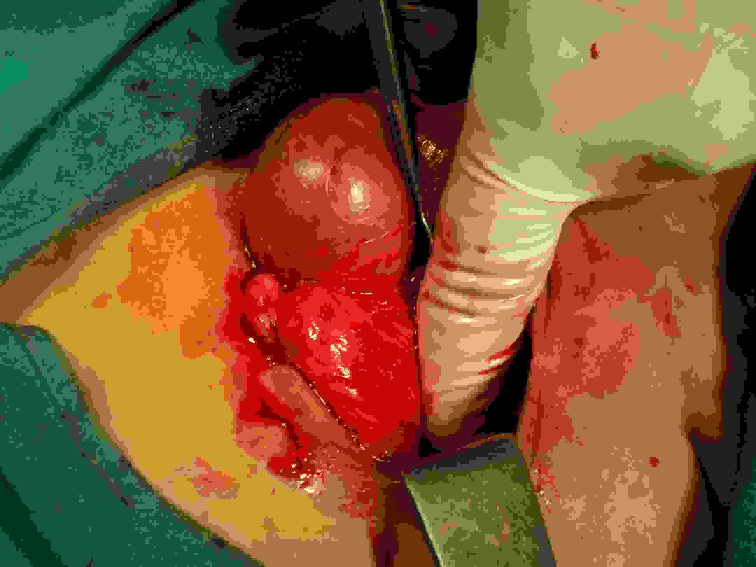
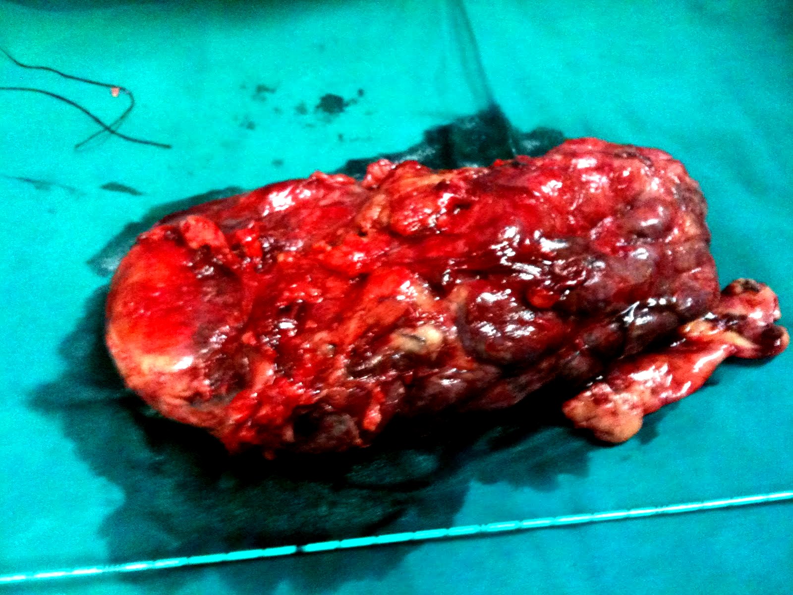
A 7–year–old boy presented with recurrent episodes of fever, dysuria and abdominal colic on and off since past 5 months. An abdominal examination revealed a lump which occupied right iliac and hypogastric areas, which measured 10×5 cm. Scrotal sac was empty on the left side and left testis was not feeling up to the deep inguinal ring. On investigating, haemoglobin was found to be 13.4 gm%, WBC count was 9400/mm3, serum Creatinine was 1.3mg%, and urine routine examination showed 8-10 pus cells. Ultrasonography of the abdomen and pelvis showed an empty left renal fossa with visualization of a gross hydronephrotic kidney, which had occupied right iliac fossa and hypogastric region. X–ray IVP showed non delineation of the left kidney [Table/Fig-1]. A 99 mTc DTPA scan was performed to assess exact kidney function and its location [Table/Fig-2]. DTPA scan revealed an unfused crossed ectopia of left kidney to the right, with a grossly reduced GFR of 7.6 ml/min. The decision to perform an open nephrectomy of the ectopic left kidney was taken, based on DTPA scan findings. At surgical exploration, left kidney was found to be ectopic and grossly hydronephrotic [Table/Fig-3a and 3b], with its ureter entering the bladder on the orthotopic site. There was a clear plane of separation between the two kidneys which were placed on right side. Left testicle was searched for its intra–abdominal location, but it could not be visualized, except for blind ending vessels on the left side. The patient had a smooth post-operative recovery, and he was asymptomatic during his follow-up. Pathological examination revealed a paper thin parenchyma on gross, and features of chronic pyelonephritis on microscopy.
Non delineated left kidney with normal opacification of the right pelvicalyceal system
DTPA scan depicting crossed renal ectopia left to right with marked reduction of GFR of ectopic moiety
Intra–operative view showing grossly hydronephrotic, ectopic kidney
Discussion
Crossed renal ectopia was first described by Pannorlus in 1964, which was defined as transposition of a kidney to the contralateral side, with midline crossing the associated ureter to be inserted in its normal position in the bladder [2,3]. McDonald and McClellan classified crossed renal ectopia into 4 types: type A, a crossed ectopia with fusion; type B, a crossed ectopia without fusion; type C, a solitary crossed ectopia and type D, a bilaterally crossed ectopia [4]. Reported autopsy incidence of fused crossed renal ectopia is 1:7500, an incidence which is ten times higher than that of unfused variety [3]. Our patient had the uncommon type B variety. Factors which are responsible for cross renal ectopia still remain undetermined. In 1938, Wilmer suggested that a crossover occurs as a result of pressure from abnormally placed umbilical arteries, that prevents a cephalad migration of the renal unit, which then follows the path of least resistance to the opposite side [5]. Although most of the individuals are asymptomatic,1/3 of cases have abdominal masses [6]. The highest incidence of associated anomalies is seen in type C. Fifty percent of patients with solitary crossed renal ectopias have skeletal anomalies, and 40% have genital abnormalities. The most common of the latter in males is either cryptorchidism or absence of the vas deferens. However, incidence of concomitant anomalies in other varieties of crossed ectopias is low; among which imperforate anuses (4%), orthopaedic anomalies (4%), skeletal abnormalities and septal cardiovascular defects are more frequent [7,8]. In our case, the left testis was absent, which per se, is a very rare association with an unfused crossed ectopia. We performed a nephrectomy, because the ectopic moiety was poorly functioning (GFR-7.6 ml/min). A 99 mTc DTPA scan is a useful tool for evaluation of a nonfunctioning ectopic kidney [5]. CECT can be used as an alternative to a DTPA scan in diagnosing renal ectopia [8]. In this case, diagnosis was made by USG, IVP and DTPA scanning. Radionuclide scanning is a must while a nephrectomy is considered for a non functioning ectopic kidney.
Conclusion
Congenital renal anomalies should be suspected in previously asymptomatic patients who present with abdominal lumps or colic. Even though they are not common, the ectopic kidney varieties should be thought of. Our finding of a cross renal ectopia (left to right) without a fusion itself was a rare occurrence and its coexistence with anorchia was again an even rarer association.
Abbreviation: USG-ultrasonography, IVP-intravenous pyelography, DTPA-diethylenetriaminepentaacetic acid, CECT-contrast enhanced computed tomography.
[1]. Shapiro E, Bauer SB, Chow JS, Anomalies of the Upper Urinary Tract. In: Wein AJ, editorCampbell-Walsh Urology 2010 10th editionPhiladelphiaW.B. Saunders:3140-41. [Google Scholar]
[2]. Birmole BJ, Borwankar SS, Vaidya AS, Kulkarni BK, Crossed renal ectopiaJ Postgrad Med 1993 39:149-51. [Google Scholar]
[3]. Felzenberg J, Nasrallah PF, Crossed renal ectopia without fusion associated with hydronephrosis in an infantUrology 1991 38:450-52. [Google Scholar]
[4]. Turkvatan A, Olcer T, Cumhur T, Multidetector CT Urography of renal fusion anomaliesDiagn Interv Radiol 2009 15:127-34. [Google Scholar]
[5]. Belekar DM, Dewoolkar V, Desai AA, Anam JR, Parab MB, An Unusual Case of non functioning Crossed Renal Ectopia without fusionThe Internet Journal of Surgery 2009 19(2) [Google Scholar]
[6]. Gopaldas RR, Walden TB, Ovulatory dysuria: a bizarre presentation of crossed non-fused ectopic kidney with extra renal pelvisInternational Urology and Nephrology 2008 40(4):889-92. [Google Scholar]
[7]. Nursal GN, Buyukdereli G, Unfused renal ectopia: a rare form of congenital renal anomalyAnnals of Nuclear Medicine 2005 19(6):507-10. [Google Scholar]
[8]. Ramaema DP, Moloantoa W, Parag Y, Crossed Renal Ectopia without Fusion—an Unusual Cause of Acute Abdominal Pain: A Case ReportCase Reports in Urology:2012 [Google Scholar]