Dermatofibrosarcoma Protuberans – A Recurrent Lesion with Unusual Presentation in the Parotid Region
Usha Hegde1, Sujeeth K Shetty2, Huchanahalli Sheshanna Sreeshyla3, Vidya Gowdappa Doddawada4
1Professor, Department of Oral Pathology and Microbiology,JSS Dental College and Hospital, Mysore, Karnataka, India.
2Professor, Department of Oral Maxillofacial Surgery,JSS Dental College and Hospital, Mysore, Karnataka, India.
3Lecturer, Department of Oral Pathology and Microbiology,JSS Dental College and Hospital, Mysore, Karnataka, India.
4Reader, Department of Oral Pathology and Microbiology,JSS Dental College and Hospital, Mysore, Karnataka, India.
NAME, ADDRESS, E-MAIL ID OF THE CORRESPONDING AUTHOR: Dr. Usha Hegde, Professor, Department of Oral Pathology & Microbiology JSS Dental College & Hospital, Mysore, Karnataka, India.
Phone: 9844453444,
E-mail:jssop12@gmail.com
Dermatofibrosarcoma protuberans (DFSP) is a rare cutaneous malignant neoplasm consisting of relatively monomorphous mononuclear spindle cells that show diffuse CD34+ positivity. Management of this lesion poses problem because of its infiltrative margins and high propensity for local recurrence and rare distant metastasis. It is said that clear margins with minimal removal of normal tissues can be achieved by Mohs’ micrographic surgery, which is particularly important in head and neck locations where the presence of vital anatomical structures and aesthetic considerations hinders a wide surgical excision. A case of DFSP, at a very uncommon site of parotid region, which recurred within 9 months after initial surgical treatment, is being reported.
Dermatofibrosarcoma protuberans, Storiform pattern, CD34+, Mohs’ surgery, Recurrence
Case Report
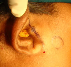
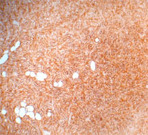
A 42-year-old female patient reported with a complaint of slow growing mass since 4 years in front of the right ear. On inspection, an inconspicuous mass was seen. It was nodular measuring about 2cm x 2cm, on palpation [Table/Fig-1]. The ultrasound scan report suggested an irregular hypertrophic soft tissue mass in the subcutaneous tissue, anterior to the parotid gland. An excisional biopsy suspecting a benign cutaneous tumor was done. The lymphnodes were not involved. The histopathological report revealed a highly cellular stroma consisting of plump monomorphic spindle cells with vesicular nuclei arranged in a storiform pattern [Table/Fig-2]. Few cells showed anisocytosis and mitotic figures less than 5 per 10 HPF. The cells were invading the adjacent adipose tissue and there was no clear demarcation of the margins. The IHC finding of diffuse positivity of tumor cells with CD34+ helped in arriving at the final conclusive diagnosis of DFSP [Table/Fig-3]. Since there was only primary tumor with no involvement of regional lymphnodes, it was categorized as stage I. In the present case the patient was treated initially by surgical excision of the lesion. Regular follow-up revealed recurrence, with the patient reporting two painful nodular swellings in the same region after 9 months. Regional and distant metastasis was ruled out. The tumor was surgically excised with negative surgical margins and was referred for adjuvant radiotherapy.
Clinical photograph showing the inconspicuous mass in front of the right ear (marked with surgical ink)
Photomicrograph showing storiform pattern of spindle cells with few mitotic figures (H < E Staining, X400)

Photomicrograph showing diffuse CD34+ positive staining tumor cells (IHC Staining, X400)
Discussion
DFSP has been reported in literature as early as 1890, but it was Hoffman who officially coined the term “dermatofibrosarcoma protuberance” [1]. It typically occurs in early or mid adult life with a slight predilection for occurrence in males and on the trunk and proximal extremities. It is reported to occur rarely in children and on uvula and parotid region [2]. DFSP begins usually as a plaque, which grows slowly over a period of several years and can manifest later as multiple small subcutaneous nodules. Hence, the “protuberant” appearance can be appreciated only in the fully developed lesions. Histopathologically, DFSP is poorly circumscribed with tumor cells infiltrating diffusely into dermis and subcutis. The tumor cells are composed of uniform population of monomorphic spindle cells arranged in storiform pattern with mitotic figures not exceeding 5 per 10 HPF [2]. It is important to distinguish this tumor from fibrous histiocytoma which is a benign tumor, since both present similar clinical and histopathological findings. However, the management and prognosis differs as DFSP is locally aggressive and known to recur in about 50% of patients and also show rare distant metastasis [3]. A positive CD34+ IHC staining of tumor cells confirms the diagnosis of DFSP and excludes tumors such as fibrous histiocytoma [3], myxoid liposarcoma and myxofibrosarcoma [4] which can mimic DFSP. The neural tumors can be excluded based on their positive IHC staining with S100protein and negative CD34+ staining [3]. Ugurel et al., have proposed a staging system according to the German Guidelines for DFSP. In this system Stage I represents the primary tumor, Stage II describes DFSP with regional lymph node metastasis and Stage III characterizes distance metastases [5].
DFSP is a fibrohistiocytic tumor of low to intermediate malignancy, having infiltrative margins with high local recurrence (about 50%) and rare distant metastasis [3]. It occurs typically in early or mid adults on the trunk and proximal extremities with slight male predilection. Its occurrence in children and on uvula and parotid region is very unusual and rare [5]. The reciprocal translocation between chromosome 17 and 22 causes fusion of collagen 1-alpha-1 gene with platelet derived growth factor B gene resulting in a potent mitogen for connective tissue cells which triggers the proliferation of DFSP tumor cells [2].
In literature, it is reported that wide excision of 3cm margins decreases the rate of recurrence significantly [6]. However, Mohs’ micrographic surgery which focuses on minimal removal of normal tissues also shows low recurrence. This is of particular significance in head and neck region [7]. DFSP is a rare locally aggressive dermal malignancy. Its occurrence in the parotid region as in present case poses great difficulty in its management due to the critical structures and esthetic difficulties in reconstruction. Although surgery is the gold standard in its treatment, adjuvant radiotherapy has been suggested in recurring lesions, when the margins are positive or close to the tumor or in cases where wider excision is not possible due to anatomic limitations, especially in head and neck regions [8]. Even chemotherapy with imatanibmesylate for advanced and distant metastatic lesions has been suggested [9].
In the present case, the rarity of location of the lesion ruled out the possibility of considering DFSP in the clinical differential diagnosis. However, correlation of the clinical presentation with histopathological findings helps in arriving at the diagnosis. Using the immunohistochemical studies a definitive diagnosis of DFSP was established. Due to the recurrence of the lesion, an adjuvant radiotheraphy has been advised in addition to the surgical management.
Acknowledgement
We wish to acknowledge Dr. Lakshman K for his help and assistance with this case.
[1]. D Lemm, LO Mugge, T Mentzel, K Hoffken, Current treatment options in dermatofibrosarcoma protuberans.J Cancer Res Clin Oncol 2009 135(5):653-65. [Google Scholar]
[2]. HB Taylor, EB Helwig, Dermatofibrosarcoma protuberans: a study of 115 casesCancer 1962 :15-717. [Google Scholar]
[3]. SW Weiss, JR Goldblum, Enzinger & Weiss`s soft tissue tumors.5th EditionChinaMosby Elsevier::371-402. [Google Scholar]
[4]. CDM Fletcher, Diagnostic Histopathology of Tumors23rd EditionChinaElsevier publications::1485-90. [Google Scholar]
[5]. N Angouridakis, P Kafas, W Jerjes, S Triaridis, Dermatofibrosarcoma protuberans with sarcomatous transformation of the head and neck. Head & Neck Oncology 2001 :3-5. [Google Scholar]
[6]. BR Burkhardt, EH Soule, RK Winkelmann, Dermatofibrosarcoma protuberans. Study of 56 cases.Am J Surg 1996 111:638-64. [Google Scholar]
[7]. HM Gloster, KR Harris, RK Roenigk, A comparison between Moh`s micrographic surgery and wide surgical excision for the treatment of dermatofibrosarcoma protuberans.J Am Acad Dermatol. 1996 :35-82. [Google Scholar]
[8]. H Suit, I Sprior, HJ Mankin, Radiation in management of patients with dermatofibrosarcoma protuberans.J Clin Oncol. 1996 :145-2365. [Google Scholar]
[9]. GA Mc Arthur, Molecular and clinical analysis of advanced dermatofibrosarcoma protuberans treated with imatinib: Imatinib target exploration consortium study.J Clin Oncol 2005 :23-866. [Google Scholar]