Anaesthetic Implications and Management of a Giant Ovarian Cyst
Nalini KB1, Prathima PT2, Shivakumar Shivanna3, Mohan CVR4
1 Assistant Professor, Department of Anaesthesiology, Critical Care and Pain, M S Ramaiah Medical College and Hospital, Bangalore, India.
2 Senior Resident, Department of Anaesthesiology, Critical Care and Pain, M S Ramaiah Medical College and Hospital, Bangalore, India.
3 Professor, Department of Anaesthesiology, Critical Care and Pain, M S Ramaiah Medical College and Hospital, Bangalore, India.
4 Professor, Department of Anaesthesiology, Critical Care and Pain, M S Ramaiah Medical College and Hospital, Bangalore, India.
NAME, ADDRESS, E-MAIL ID OF THE CORRESPONDING AUTHOR: Dr Shivakumar Shivanna, Professor, Department of Anaesthesiology, Critical Care and Pain, M S Ramaiah Medical College and Hospital, Bangalore-560043, Bangalore, India.
Phone: 0919611988538,
E-mail: shivanna03@gmail.com
With improvement in health care, improved access to hospitals and better imaging modalities, huge abdominal tumours are rarely seen in modern day surgical practice. They present many challenges to anaesthesiologists. Difficult intubations, life threatening cardiovascular and pulmonary complications, are commonly encountered. Management of such cases is associated with significant mortality and morbidity. The consequences of surgery are mainly attributable to the size of the mass rather than to its distinctive pathology. We hereby report a very rare case of a giant ovarian cyst weighing 57kg, which was successfully managed by a careful pre-operative evaluation, maintenance of intraoperative haemodynamic and fluid management.
Giant ovarian cyst, Anaesthetic implications
Case Report
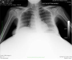
A 50-year-old female presented with an abdominal swelling which gradually increased in size over the past 13 years, due to which she was unable to ambulate. She weighed 90kg, with a circumferential abdominal girth which measured 170 cms. The umbilicus was stretched, with engorged veins all over the abdomen. The patient became dyspnoeic on lying supine. Blood investigations revealed hypochromic microcytic anaemia (Hb 7.1 gm %, PCV of 24.9). Other blood investigations were normal, with the exception of low serum albumin levels. Electrocardiogram (ECG), echocardiography (ECHO), and arterial blood gases (ABG) were within normal limits.Pulmonary function tests showed FVC-87%, FEV1-115%, FEV1/FVC -121%. Her chest X-ray showed a bilateral upward diaphragmatic displacement with normal lung fields [Table/Fig-1]. Computerized tomography (CT) of abdomen showed a huge cystic mass which arose from the right ovary, and occupied the whole abdominopelvic space [Table/Fig-2]. The patient was scheduled for an exploratory laparotomy, with excision of cyst. Pre-operatively, adequate packed cells, fresh frozen plasma, and platelets were arranged. The patient was premedicated with Pantoprazole, Ondansetron and Glycopyrrolate, 60 minutes prior to the surgery.
Chest X-ray showing displacement of the diaphragm
Giant ovarian cyst which was surgically removed and CT scan abdomen showing giant ovarian cyst

In view of respiratory distress seen while patient was in the supine position, left lateral position was inevitable for anaesthesia and surgery. An extension was applied to the operating table, to accommodate the tumour. In the theatre, ECG, saturation (SPO2), non-invasive blood pressure (NIBP) monitoring was instituted and a 16 G peripheral intravenous line was secured. A rapid sequence intubation was planned and after adequate preoxygenation, the patient was induced with midazolam, fentanyl and thiopentone. Suxamethonium 50mg was administered to facilitate endotracheal intubation. Airway was secured with a 7mm oral cuffed endotracheal tube (ETT) and anaesthesia was maintained with nitrous oxide: oxygen mixture and 0.5-1% isoflurane. Long acting muscle relaxation was provided with atracurium, based on train of four (TOF) responses. Right internal jugular vein and left radial artery was cannulated for invasive blood pressure monitoring. Continuous ECG, SPO2, heart rate, invasive blood pressure, CVP, urine output, nasopharyngeal temperature and blood loss were monitored.
Surgery was performed in the left lateral position and encysted mass was removed in total, which weighed 56.9 kg. Following the removal of the mass, there was a sudden fall in blood pressure to 70 mmHg systolic. There was considerable blood loss from the abdominal wall, with an estimated blood loss of 900 ml. The patient was resuscitated with crystalloids, 3 units of packed cells, and fresh frozen plasma. As the blood pressure did not improve despite fluid resuscitation, dopamine was started at15 mcg/kg/min, and it was then tapered to 5 mcg/kg/min after the blood pressure had stabilized. As the patient was normothermic and normotensive, she was reversed at the end of the procedure with glycopyrrolate and neostigmine and extubated. Post-operatively, the patient was transfered to the ICU for further monitoring. Intravenous fentanyl was given for post-operative analgesia. The patient remained haemodynamically stable and the dopamine infusion was tapered and discontinued on 3rdday. On the 4th post-operative day, the patient was started on oral diet.
On the 7th post-operative day, the patient developed sudden hypotension. On examination, her BP was found to be 60/36 mm Hg, CVP was 6 cm, with severe pallor. Investigations showed haemoglobin of 4gm%. Ultrasonography (USG) of abdomen showed a bilateral pleural effusion and an echogenic lesion in infraumbilical region, which were suggestive of a clot. Following her resuscitation, the patient was taken up for emergency laparotomy. The patient was induced with ketamine and atracurium and she was intubated with 7 mm ETT. Four units of packed cells were transfused intraoperatively. The bleeding was found to come from the abdominal wall, which was promptly controlled. The patient remained haemodynamically stable throughout the procedure. Post-operatively, the patient was extubated and she was shifted to ICU. On the 3rd post-operative day, the patient was ambulated with support and she was discharged from the ICU and subsequently from the hospital. Her post-operative weight was 32 kg. Histopathology revealed the mass to be mucinous cystadenoma of ovary.
Discussion
As large abdominal tumours rarely present in this era, due to greater health awareness and access to medical care, it is unlikely that many clinicians have experience in dealing with such cases. The two most common types of ovarian neoplasm are serous [1, 2] and mucinous cystadenomas, with mucinous type accounting for 15% of all ovarian tumours [3]. The potential complications of these cysts are torsion, haemorrhage and rupture. Surgical management of cysts is done by laparotomy or laparoscopic cyst excision [4].
Huge abdominal tumours interfere with respiratory function by producing elevation and splinting of the diaphragm, with flaring of rib cage. This leads to marked dyspnoea and patients are unable to lie supine. Large abdominal tumours cause supine hypotension syndrome due to aortocaval compression [5]. In these patients, the blood pressure is kept stable by maintaining a balance between the reduced cardiac output and peripheral vasoconstriction. Sympathetic blockades caused by central neuraxial blocks can abolish this protective mechanism, thus causing severe hypotension and hence, spinal or epidural anaesthesia is preferably avoided. Nevertheless, few cases in which epidural anaesthesia was administered for decompression of cysts, without producing circulatory depression or pulmonary oedema, have been reported [6]. The advantage of decompression is shrinking the cyst to a more manageable size and subsequent ease of surgical removal [7].
A large bore venous access should be obtained for anticipating severe blood loss intraoperatively [7]. The severe hypotension seen in our patient could be attributed to the splanchnic shock which had occurred when the tamponade effect of the mass on the abdominal wall was removed. Hypovolaemia, coagulation abnormalities are commonly seen in the post-operative period [7]. Coagulation abnormalities may be caused by consumption and dilutional coagulopathy, which should be treated with transfusion of fresh frozen plasma and platelets. Delayed extubation is recommended, as some patients require reintubation for ventilatory failure [7]. We decided to extubate the patient as she was normothermic and normotensive and as she was making good respiratory efforts. In the early post-operative period, haemorrhage, hypothermia, pulmonary oedema which are secondary to large transfusions [8] are common. Inadequate ventilation which is secondary to pain and altered respiratory mechanics were the reasons for providing an elective ventilation [7].
Intestinal ileus and gaseous distension do occur in these cases, which can be fatal [8]. Nasogastric aspiration, maintaining electrolyte balance and use of an elastic abdominal binder can reduce these complications [7,8]. Prophylactic measures such as elastic stockings, rapid mobilization, subcutaneous heparin and dextran infusion are advised, to reduce deep venous thrombosis and pulmonary embolism complications in these patients [7].
Conclusion
Huge ovarian tumours are rarely encountered in the modern day clinical practice. Anesthesiologists must be aware about the anaesthetic challenges that are seen in these cases. A huge ovarian tumour which weighed around 57 kg was successfully removed by providing a meticulous perioperative anaesthetic management.
[1]. Vellanki Venkata Sujatha, Sunkavalli Chinna Babu, Giant ovarian serous cystadenoma in a postmenopausal woman: a case reportCases Journal 2009 2:7875 [Google Scholar]
[2]. Florent Valour, Erwan Oehler, (Images ) A giant ovarian cystBritish Medical Journal case reports 2012 10:1136 [Google Scholar]
[3]. Nwobodo EI, Giant mucinous cystadenoma: case reportNigerian Journal of Clinical Practice 2010 13(2):228-29. [Google Scholar]
[4]. Su-Kon Kim, Jong-Soo Kim, Choong-Hak Park, Jin-Wan Park, A case of giant ovarian cyst managed successfully through laparoscopic surgeryKorean J Obstet Gynecol 2012 55(7):534-37. [Google Scholar]
[5]. Kuczkowski KM, Strock D, Large ovarian cyst as a cause of aortocaval compressionAnaesthesia 2004 59:1148 [Google Scholar]
[6]. Nishiyama T, Hanaoka K, Same day drainage and removal of a giant ovarian cystCan J Anaesth 1997 44(10):1087-90. [Google Scholar]
[7]. Morrison P, Morgan G, Removal of a giant ovarian cyst. Anaesthetic and intensive care managementAnaesthesia 1987 42:965-74. [Google Scholar]
[8]. Hoile RW, Hazard in the management of large intra-abdominal tumoursAnnals of the Royal College of Surgeons 1976 58:393-97. [Google Scholar]