Chondrolipoma of the Lower Lip: A Case Report
Kamakshi G1, Yaranal PJ2
1 Assistant Professor, Department of Conservative Dentistry and Endodontics,Kannur Dental College, Kannur, India.
2Professor, Department of Pathology,Kannur Medical College, Kannur, India..
NAME, ADDRESS, E-MAIL ID OF THE CORRESPONDING AUTHOR:
Dr. Parasappa Joteppa Yaranal, Department of Pathology, Kannur Medical College, Kannur-670612, India.
Phone: 9448824484,
E-mail: pjyaranal@yahoo.co.in
A chondrolipoma is an extremely rare form of a benign mesenchymal tumour which contains mature fatty tissue and cartilage. We are presenting a case of chondrolipoma of the lower lip which was seen in a 6-year-old girl. Chondrolipomas are rare neoplasms; their terminologies and pathogeneses have been controversial in the past. Chondrolipomas are uncommonly seen in the oral cavity, in children and in females. Hence, we are reporting this present case because rarity of this lesion.
Chondrolipoma, Lower lip, Oral lipoma
Case Report
A 6-year-old girl presented with a painless swelling in the lower lip. It had been present since the past one year, without any history of trauma. Clinical examination confirmed a sessile soft-to-firm swelling which measured 1.5 cm × 1cm, in the left side of the lower lip. No other lesions were observed in the oral cavity and tumour was well-defined from surrounding structures. An excision biopsy was done, followed by a histopathological examination.
Morphology
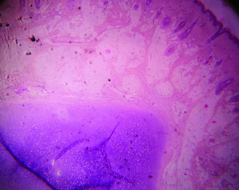
On gross examination, the tumour was found to be 1 cm x 0.5 cm in size. Cut surface showed a well encapsulated lesion which was composed of fatty tissue, with scattered plump white areas. Histopathological examination revealed a well delineated tumour which was composed of proliferating mature adipocytes, which were arranged in lobules, which were separated by thin fibrovascular septae. These lobules were interspersed with lobules of mature cartilage [Table/Fig-1,2]. Lipocytes and chondrocytes lacked any mitotic activity or nuclear atypia. No myxoid areas and no lipoblasts were detected. A diagnosis of a chondrolipoma was made.
Discussion
A chondrolipoma is an extremely rare, lipomatous tumour of benign soft tissue, which contains mature fatty tissue and mature cartilage. Chondrolipomas may be found almost anywhere in the body, particularly in the connective tissues of the breast, head and neck area, as well as in the skeletal muscle. Its different histopathological variants have been recognized, such as fibrolipoma, angiolipoma, myolipoma, spindle cell lipoma, chondroid lipoma and osteolipoma.[1,2] .
Lipomas are one of the most common mesenchymal tumours which constitute 1-4% of tumours which occur in oral cavity. [3] Oral lipomas are more common in males and they are seen between the ages of 40- 60 years. Oral lipomas are slow growing neoplasms and they are relatively asymptomatic [3,4].
Stout [5] first proposed the term, ‘mesenchymoma’ to designate a benign tumour which was composed of a mixture of two or more nonepithelial elements other than fibrous tissue, but this term has now been abandoned due to lack of its specificity. Other terms like choristoma, which defines a tumour-like ectopic rest of normal tissue, and hamartoma, which designates an overgrowth of mature tissues, which are normally present at the affected site, have also been proposed [6] . At present, the most established opinion is that these lesions are true neoplasms (lipomas) with cartilaginous metaplasia and that they should be best referred to as chondrolipomas [1,7] .
The pathogenesis of chondrolipoma is unknown, but two possible explanations have been proposed for cartilage and bone formations in benign mesenchymomas [3,4] . The first is that cartilage arises from chondro-osseous metaplasia of adipose tissue, presumably due to mechanical stress or trophic disturbance [1] . The close contact or proximity of tumours to bone or large joints is often associated with metaplastic changes [8] . The second is that the cartilage may originate from differentiation of multipotential cells in the mesenchymomas. A recent study reported abnormal expressions of transforming growth factor-ß (TGF-ß), latent TGF-ß binding protein-1 (LTBP-1) and bone morphogenetic protein (BMP) in chondrolipomas, which pointed to unique pathogenesis of this neoplasm [8] . Considering the proximity of chondrolipomas to mandibular bone, as it was in our case, microtrauma may the cause of the chondro-osseous metaplasia.
Chondrolipomas have no specific radiographic features. The diagnosis is based on a histopathological examination. Histologically, chondrolipomas are characterized by lobules of mature adipocytes with islands of mature cartilaginous tissue [1,4] . In our case, similar histomorphological features were identified. Because morphological differential diagnoses are lacking, making a histological diagnosis of a chondrolipoma is straight forward. However, clinicians and pathologists should be aware of the terminological (but not histological) similarity between chondrolipomas and chondroid lipomas. The latter term defines a unique and a recently recognized benign adipose tissue tumour which contains a chondroid matrix, fat and lipoblasts and which therefore, resembles a myxoid liposarcoma or an extraskeletal myxoid chondrosarcoma [1] .
Despite the controversies which are existent in the pathogeneses of chondrolipomas of the oral cavity, the treatment of choice is complete surgical excision. No data on recurrence rates and malignant transformations of chondrolipomas are available until now [1,4] .
Chondrolipomas are uncommonly seen in the oral cavity. Clinical and histological characteristics of chondrolipomas have been poorly understood because of their rarity, together with the lack of data in the literature. The present case thus provides the spectrum of neoplasms which are known to arise at this anatomical site.
Well encapsulated lesion composed of lobules mature adipocytes separated by connective fibrous septa. (H & E x100)

Histopathological examination showed island of mature cartilage is surrounded by mature fat tissue. (H & E x400)

Conclusion
To the best our knowledge, limited number of cases (10 cases) of chondrolipomas which occurred in the lower lip has been reported. Chondrolipoma is a rare neoplasm whose terminology and pathogenesis has been described inconsistently by various authors in the past. In the English-language literature, very limited number of reported cases of chondrolipomas of oral cavity has been described. Therefore, the present case expands the spectrum of neoplasms which are known to arise in the oral cavity.
[1]. SW Weiss, JR Goldblum, Benign lipomatous tumors. In: Weiss SW, Goldblum JR, Editors. Enzinger and Weiss’s soft tissue tumors. 2001 154th edSt. LouisMosby Publication;:571-639. [Google Scholar]
[2]. RM Castilho, CH Squarize, FD Nunes, DS Pinto Junior, Osteolipoma: a rare lesion in the oral cavity.Br J Oral Maxillofac Surg 2004 42:363-4. [Google Scholar]
[3]. MA Furlong, JC Fanburg-Smith, ELB Childers, Lipoma of the oral and maxillofacial region: site and subclassification of 125 cases.Oral Surg Oral Med Oral Pathol Oral Radiol Endod 2004 98:441-50. [Google Scholar]
[4]. CFW Nonaka, MCDC Miguel, LB Desouza, LP Pinto, Chondrolipoma of the tongue: a case report. J Oral Science 2009 51:313-6. [Google Scholar]
[5]. AP Stout, HO Heymann, Mesenchymoma, the mixed tumor of mesenchymal derivatives.Ann Surg. 1948 127:278 [Google Scholar]
[6]. JR Austin, M de Tar, DH Rice, Pulmonary chondroid hamartoma presenting as an inflatable neck mass. Case report and clinicopathologic analysis.Arch Otolaryngol Head Neck Surg. 1994 120:440-3. [Google Scholar]
[7]. R Ito, M Fujiwara, K Takagaki, R Nagasako, Chondrolipoma of the toe.J Dermatol. 2007 34:570-2. [Google Scholar]
[8]. M Nakano, E Arai, Y Nakajima, H Nakamura, K Miyazono, T Hirose, Immunohistochemical study of chondrolipoma: possible importance of transforming growth factor (TGF)-betas, latent TGF-beta binding protein-1 (LTBP-1), and bone morphogenetic protein (BMP) for chondrogenesis in lipoma.J Dermatol 2003 30:189-95. [Google Scholar]