Impacted Teeth: A Radiographic Pictorial Essay
Beena Varma R1, Nidhin J Valappila2, Nimisha Mathew3
1Associate Professor & Head, Department of Oral Medicine & Radiology, Royal Dental College, Chalissery, Palakkad, Kerala, India.
2Assistant Professor, Department of Oral Medicine & Radiology, Royal Dental College, Chalissery, Palakkad, Kerala, India.
3Lecturer, Department of Oral Medicine & Radiology, Royal Dental College, Chalissery, Palakkad, Kerala, India.
NAME, ADDRESS, E-MAIL ID OF THE CORRESPONDING AUTHOR: Dr. Beena Varma R, Associate Professor & Head, Department of Oral medicine & Radiology, Royal Dental College, Chalissery, Palakkad -679536, India. Phone : 8606315371, 9747672871,
E-mail: beenavarmar@gmail.com
Impaction, Radiographic essay, Radiography of impaction, Types of impaction
Introduction
Mammalian development shows increased progressive uneruption, impaction and agenesis which may lead to extinction of a few teeth. A report of a 13,000- to 15,000-year-old skeleton of a 25- to 35-year-old woman excavated in France in 1911 showing impacted wisdom teeth may contradict the theory of dietary change [1]. In 1992 the World Health Organization stated that one of the goals for the year 2020 was people with a functional and esthetic dentition of at least 20 teeth throughout their life without any dental prosthesis [2]. Impaction is the failure to erupt due to a physical barrier whereas primary retention is failure of eruption before gingival emergence without any barrier or ectopic position and secondary retention is failure to attain occlusal position without any physical barrier [3].
This article is based on a retrospective radiographic survey of few panoramic and intraoral radiographs of the archives of Royal Dental College and the author’s personal collection. Various radiographic presentations of impacted teeth are analyzed. Attempt is made only to see the types of impaction as evidenced in the radiograph and not any statistical analysis. Recently the importance of teeth as a model organ system for evolutionary studies is being highlighted [4] [Table/Fig-1,2,3,4,5,6,7,8,9,10,11,12,13,14,15,16,17,18,19,20,21,22,23,24,25,26,27,28,29,30,31,32,33,34,35].
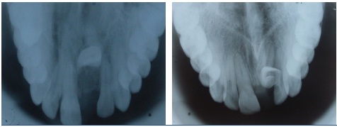
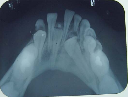
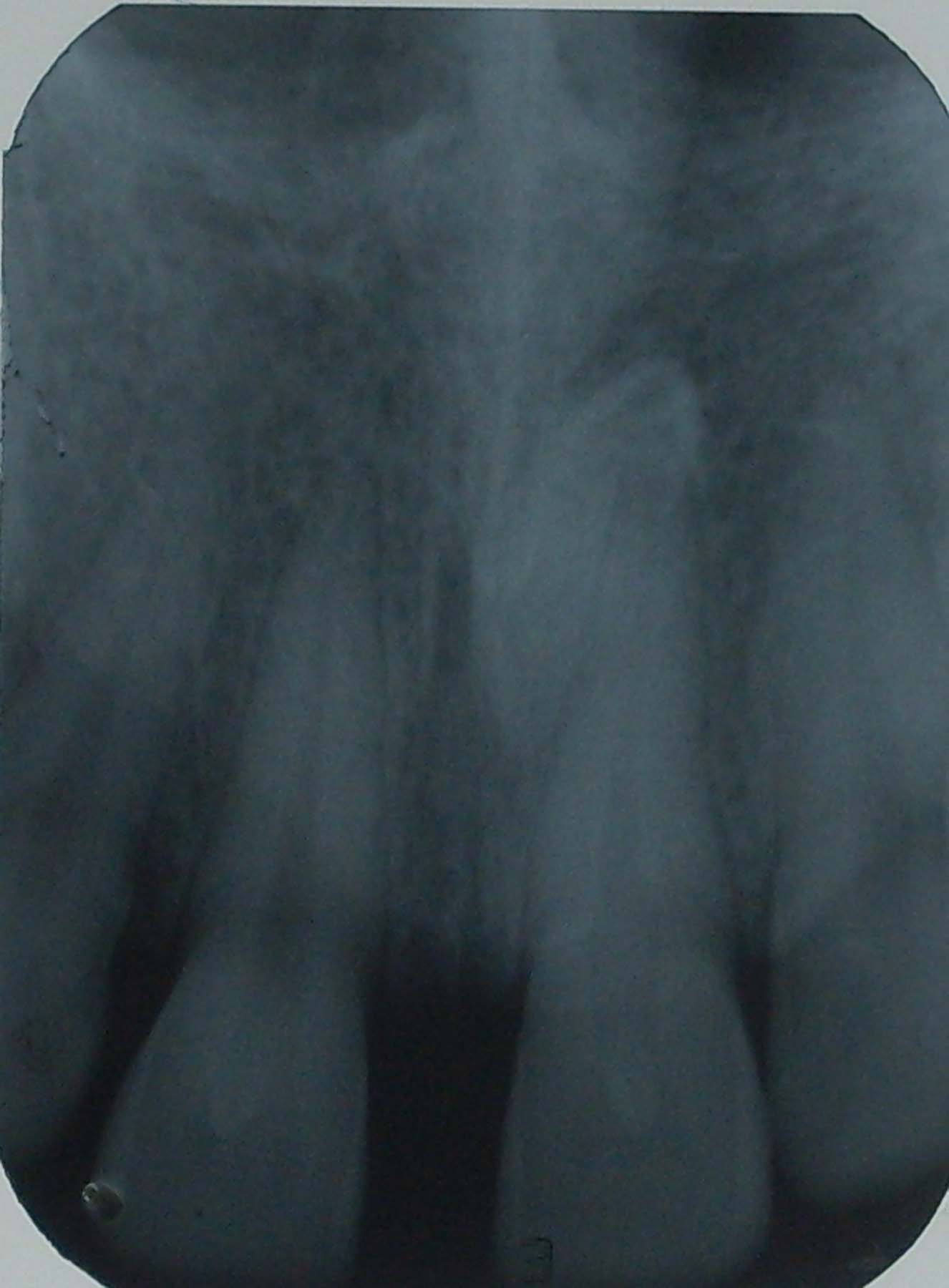
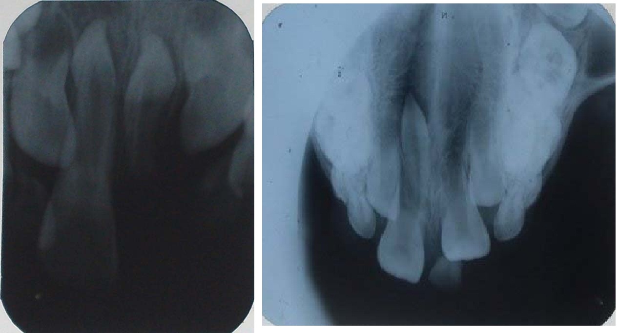
Impacted central incisors The patients gave histories of trauma of the deciduous tooth. The permanent tooth has a dilaceration at the cementoenamel junction in both cases. Similar cases are reported in literature
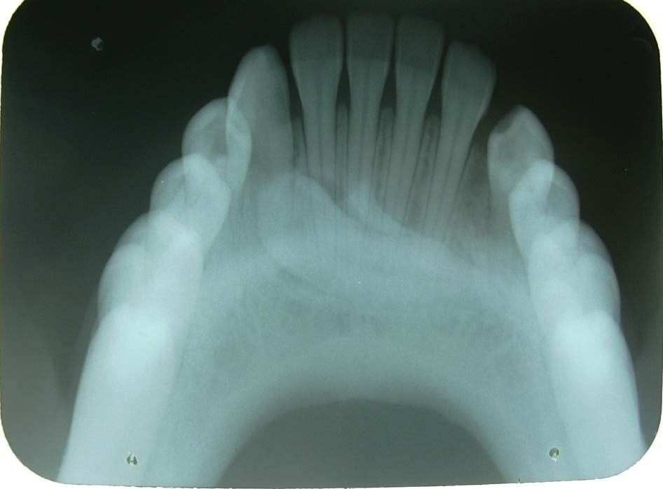
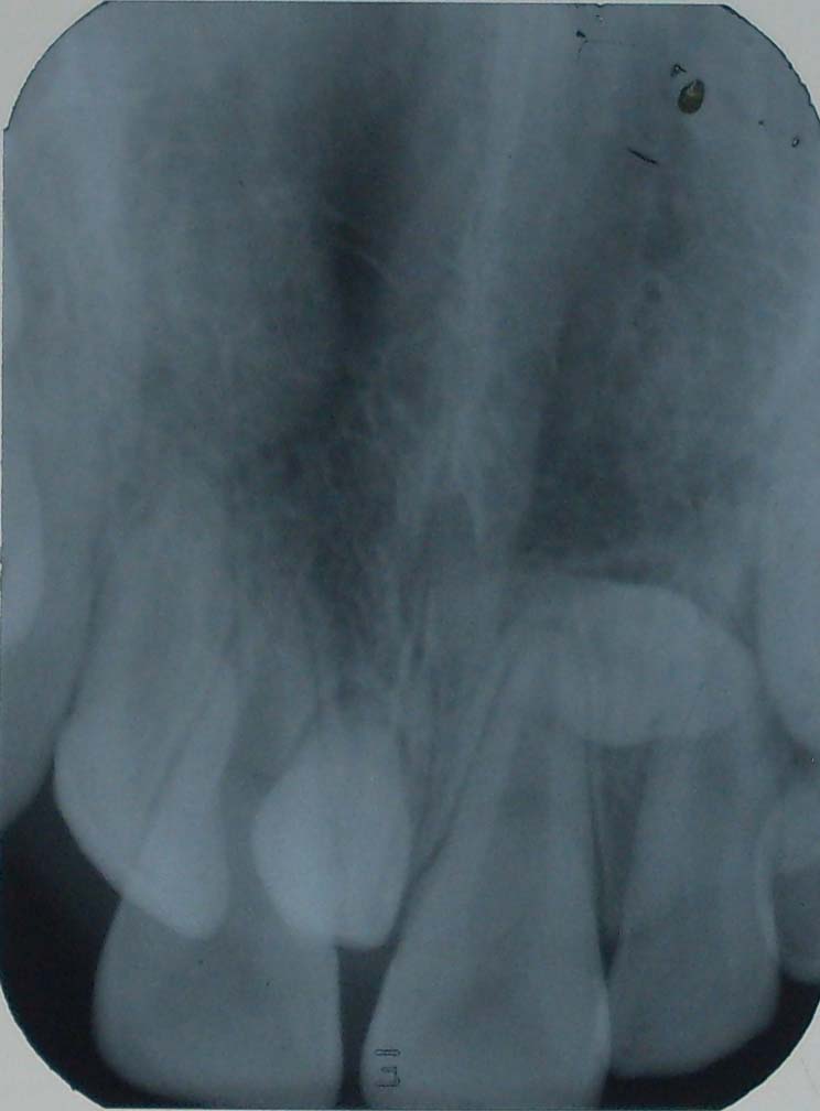
Bilaterally impacted maxillary canine with retained deciduous lateral incisors and canines

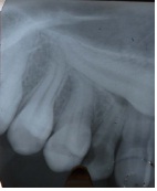
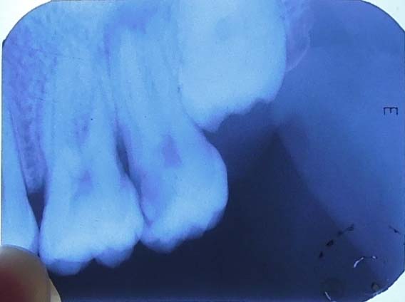
Impacted maxillary canine with deciduous canine in situ with no signs of resorption
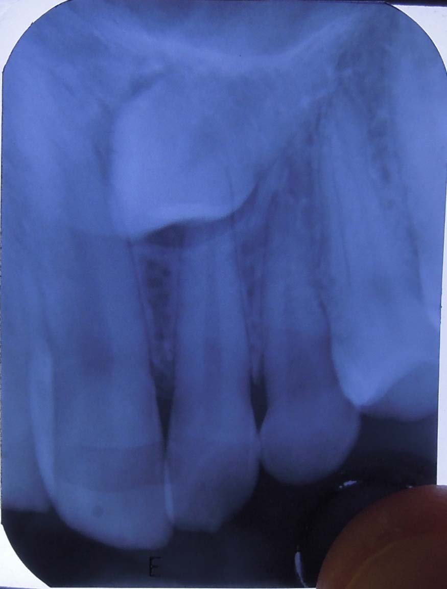
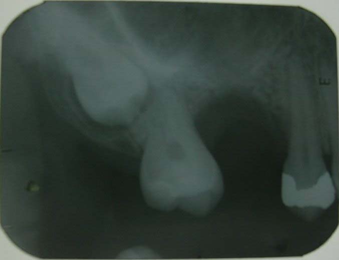
Impacted maxillary canine with minimal resorption of deciduous canine. Note the dilaceration of roots of premolars at the cervical one- third
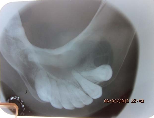
Shows horizontal impaction of mandibular right canine. Left canine has normaly erupted
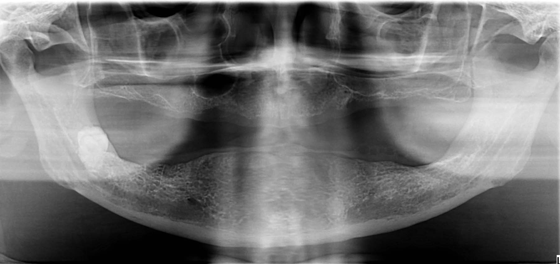
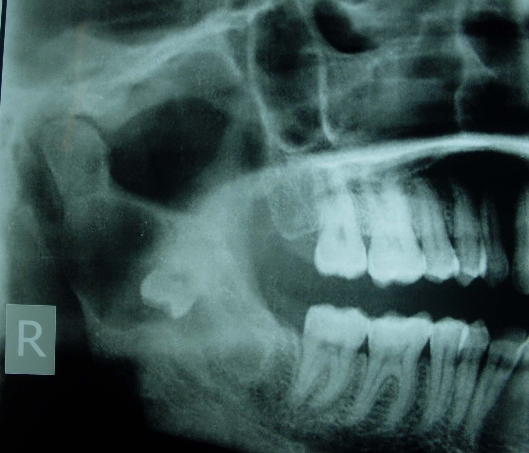
Horizontally impacted teeth obstructed by odontome
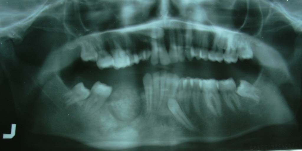
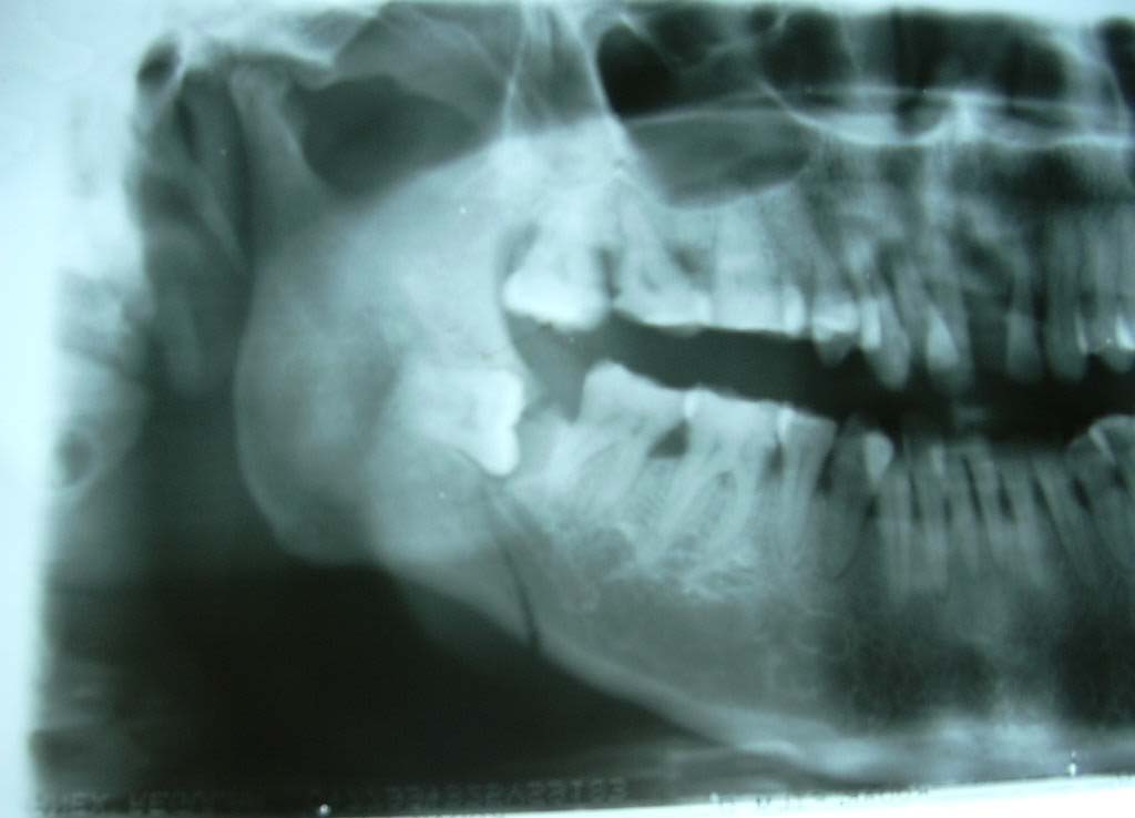
Impacted mandibular canine with associated cystic lesion
Another case of impacted mandibular canine with other anomalies like odontome and associated cystic lesion
Another case of both maxillary and madibular canine impaction with other anomalies like odontome and associated cystic lesion in the mandible
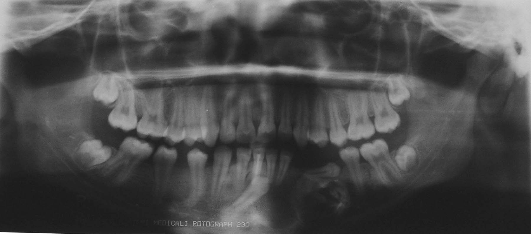
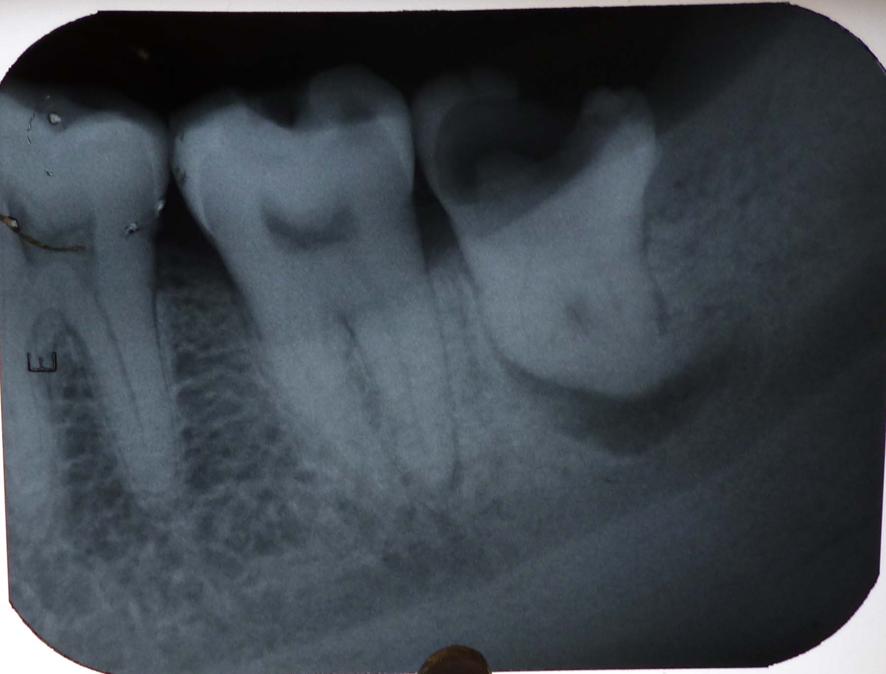
Developing maxillary third molars with taurodontism of maxillary second molars. Mandibular second molars also shows mild increase in pulp chamber. These have high chance of getting impacted. Impaction may be associated with other anomalies

Mesioangular impaction of maxillary third molar
Vertical impaction of maxillary third molar
14 Distally and horizontally impacted maxillary third molar. Most maxillary impactions may be asymptomatic or may cause TMJ problems in the long run. In this patient who presented with TMJ pain, one of the contributing factors for the TMJ changes is the impacted tooth. The impacted tooth was an incidental finding
A rare case of maxillary first molar impaction. First permanent molar usually get impacted due to ectopic eruption and this may lead to resorption of primary second molar distal root which in turn may cause root resorption and early loss [5]
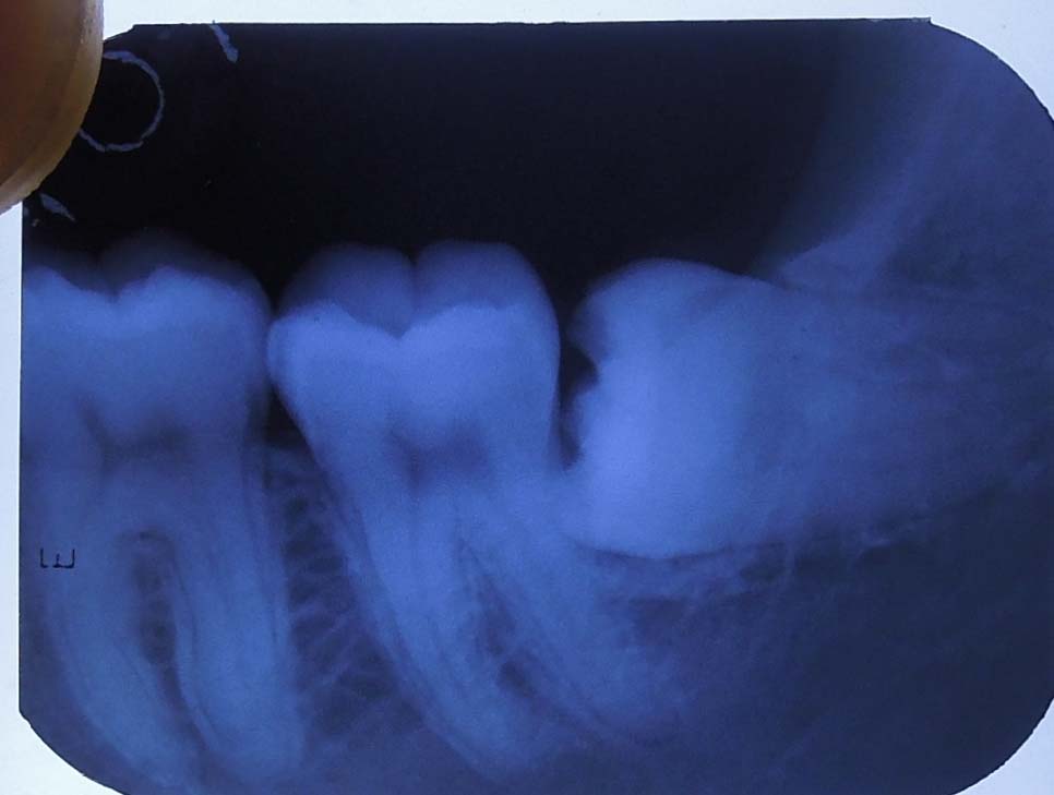
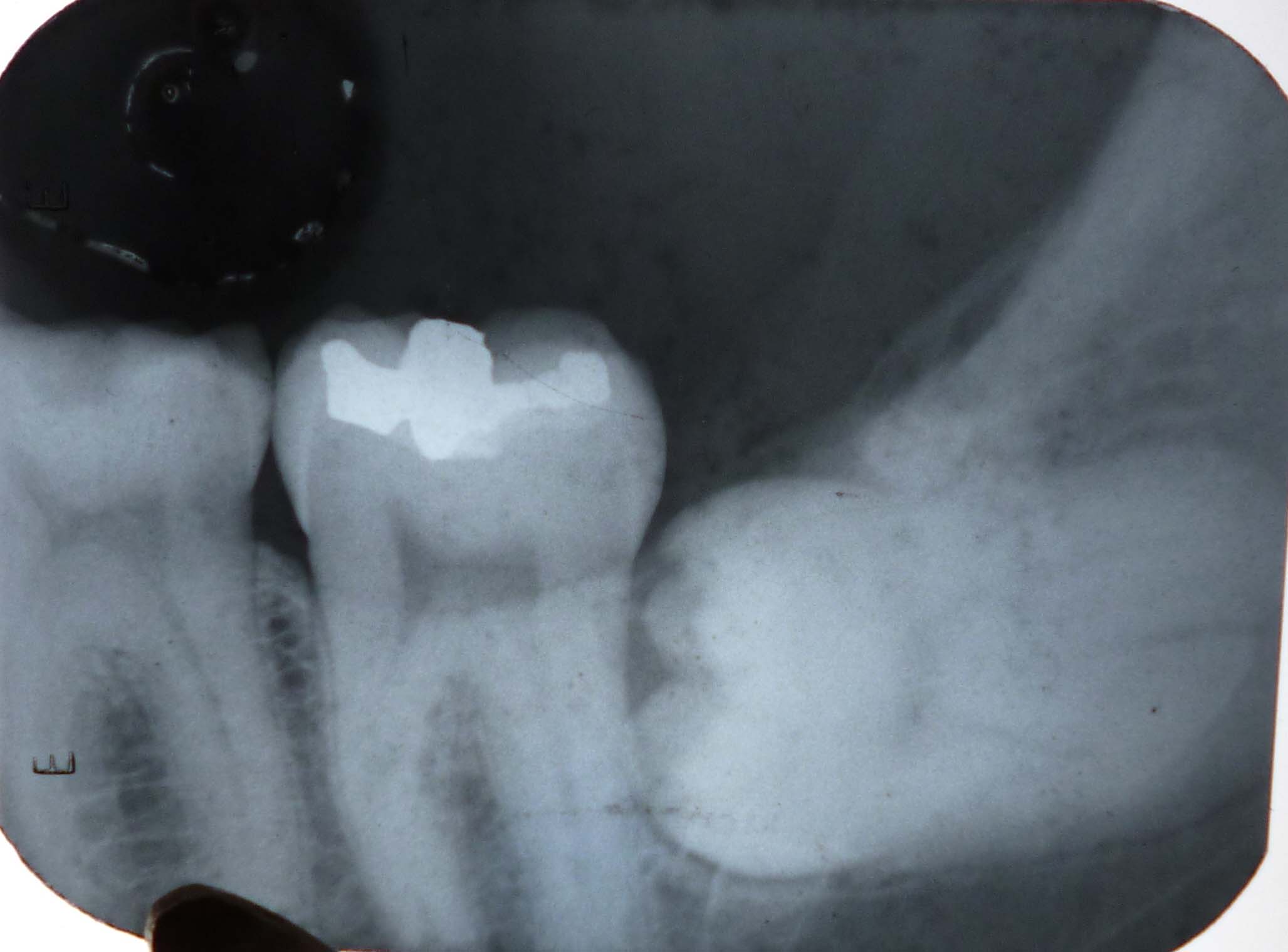
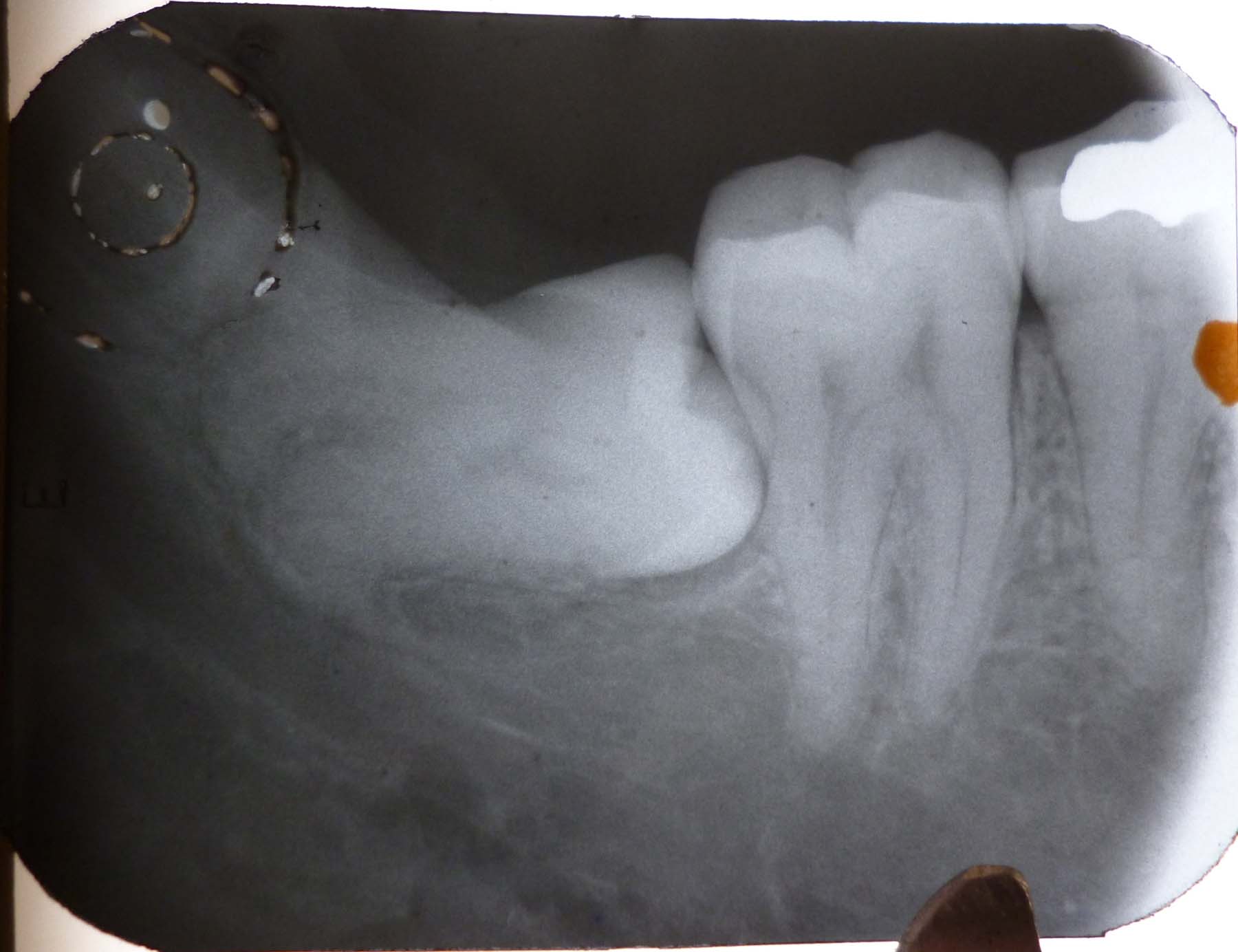
Horizontal impaction of mandibular third molar
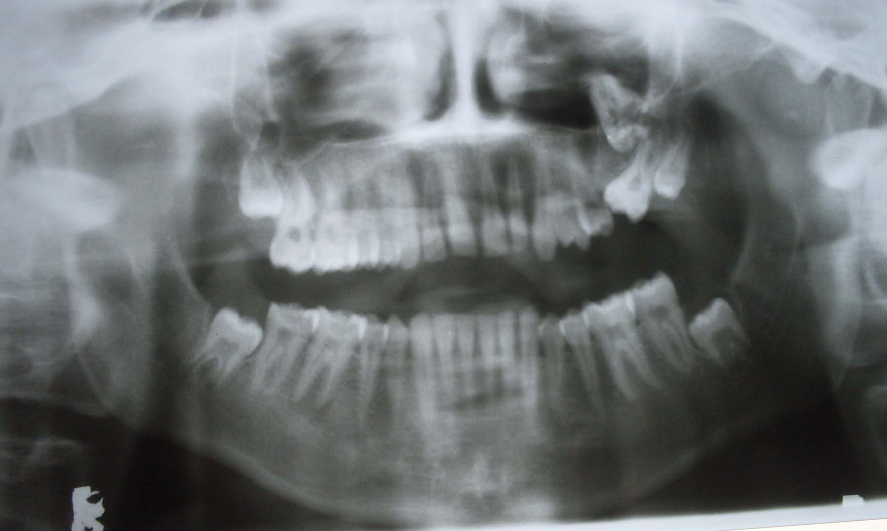
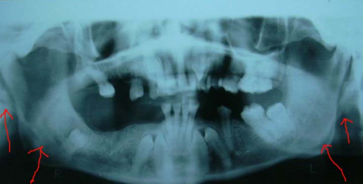
Non syndromic multiple impacted teeth -. Elongated styloid process also seen
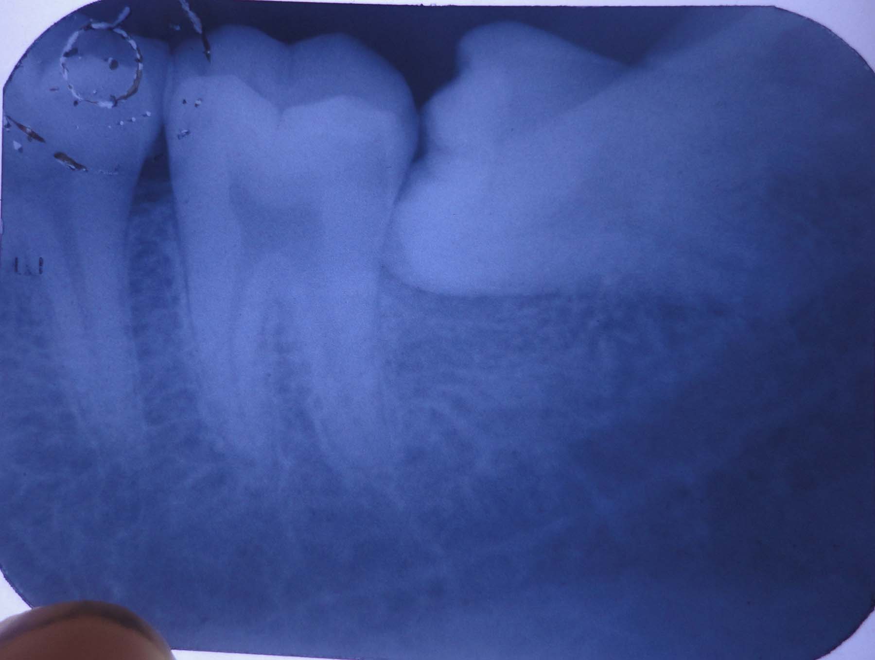
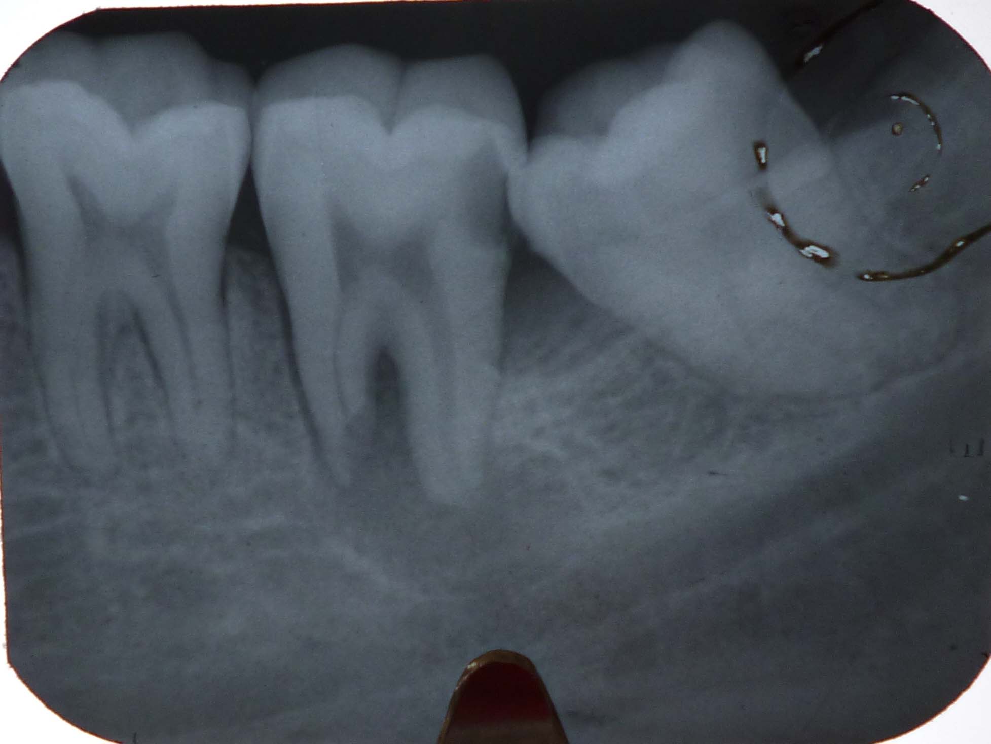
A case of impacted mandibular third molar
Vertical impactions with distally developing cystic lesion ,Fig 19 the third molar shows dilaceration also
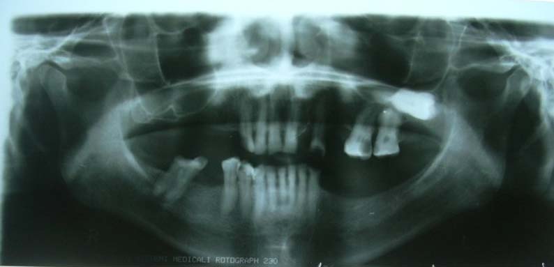
Panoramic radiograph of a patient taken before complete denture prosthesis fabrication. Impacted third molar (Pell & Grigory Class III) Patient was asymptomatic.
Impacted mandibular third molars in a buccolingual direction (Winter’s classification 5)

Cropped view of panoramic radiograph showing fracture along impacted third molar (mesioangular impaction). Impacted tooth is a weak area in the bone and Impacted third molars are susceptible areas causing mandibular angle fracture
Mesioangular impaction of third molar with an associated large radiolucent lesion and distal root resorption of second molar. They are prone to induce cystic changes
Pell & Gregory Class II impacted third molar with a radiolucent lesion extending to the neck of the condyle. The tooth is inverted and is associated with cystic degeneration of bone with extensive resorption necessitating considerable bone resection and facial deformity. Hence when suspected of impaction periodic radiographic evaluation is warranted
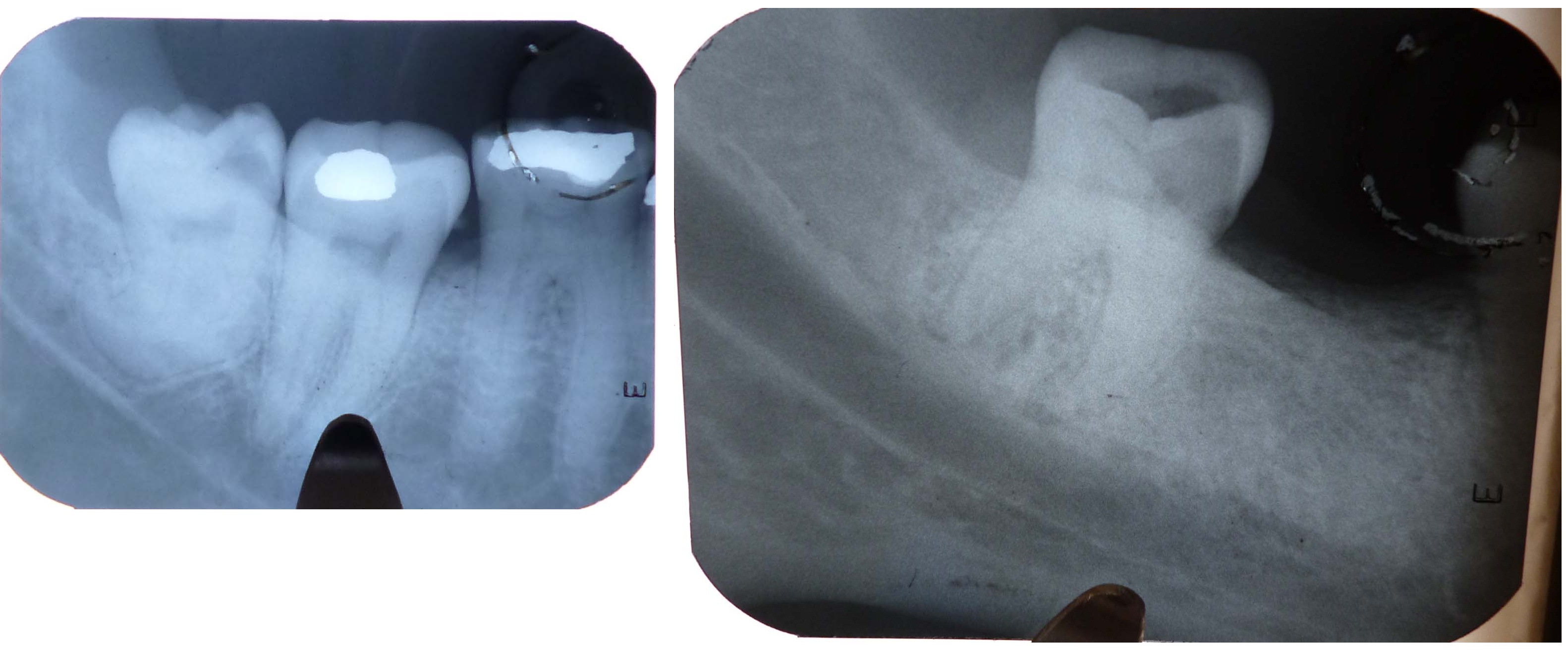
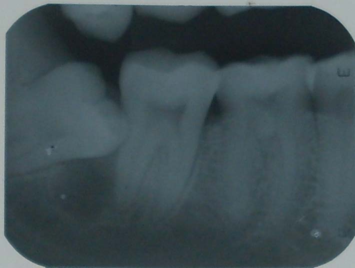
An asymptomatic horizontal impaction of third mandibular molar Surgically challenging because of its close proximity to the mandibular canal
Horizontally impacted mandibular third molar with dilacerated roots with root close to the mandibular canal. This is also a surgical challenge
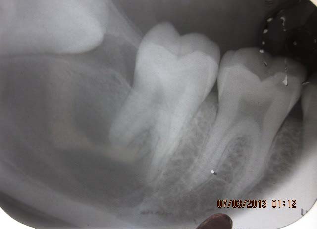
Mesioangular (placement) impaction of mandibular third molar with interdental bone resorption and root resorption of second molar. Secondmolar shows proximal caries & periapical pathology
A mesioangular impaction of mandibular third molar with a developing cystic lesion below the crown
Vertical impaction of mandibular third molar with dilaceration of root. Tooth is decayed with periapical pathology close to the mandibular canal; a challenge to the oral surgeon
Impacted inverted mesiodens
Impacted inverted mesiodens . Fig 33 shows two inverted mesiodens
Multiple impacted supernumerary teeth in the maxillary region. (Similar to Fig. 1 & 2 ) Supernumeraries may occur unassociated with syndromes
Conclusion
This article is an attempt on retrospective analysis of radiographs showing impacted tooth. The variety and types of impaction prevalent seems more than what is reported in the literature, at least in this part of the country where this analysis was done. This invites further detailed studies on the causes and frequencies of impacted tooth and the pathology associated with them.
[1]. Source: Magdalenian Girl is a woman and therefore has oldest recorded case of impacted wisdom teeth” (Press release).Field Museum of Natural History. 2006 March 7 [Google Scholar]
[2]. Hobdell Martin, Petersen Poul Erik, Clarkson John, Johnson Newell, Global goals for oral health.2020 International Dental Journal. 2003 53:285-88. [Google Scholar]
[3]. Nagpal A, Sharma G, Sarkar A, Pai KM, Eruption disturbances: an aetiologicalcum-management Perspective.Dentomaxillofacial Radiology. 2005 34:59-63. [Google Scholar]
[4]. Polly P. David , Development and evolution occlude: Evolution of development in mammalian teeth.PNAS 2000 97(26):14019-21. [Google Scholar]
[5]. BC Kirtaniya, V Sachdev, A Sachdev, P Sachdev, Management of Impacted Permanent First Molar- A Case Report.Indian Journal of Dental Sciences. 2010 2(3):p27 [Google Scholar]