Avascular Necrosis of Femoral Head in a Child with Beta Thalassaemia Major
Adharsh Narain Thulasidhar1, Sandeep Kumar2, Shrikiran Aroor3, Suneel Mundkur4
1 Senior Resident, Department of Paediatrics, Kasturba Medical College, Manipal, Karnataka, India.
2 Assistant Professor, Department of Paediatrics, Kasturba Medical College, Manipal, Karnataka, India.
3 Professor, Department of Paediatrics, Kasturba Medical College, Manipal, Karnataka, India.
4 Professor, Department of Paediatrics, Kasturba Medical College, Manipal, Karnataka, India.
NAME, ADDRESS, E-MAIL ID OF THE CORRESPONDING AUTHOR: Dr. Sandeep Kumar, Assistant Professor, Department of Paediatrics, Kasturba Medical College, Manipal University, Manipal-576104, India.
E-mail: bksandydoc@gmail.com
Dear Editor
Avascular Necrosis (AVN) of the head of femur is well known to occur in haematologic disorders associated with thrombosis. Sickle cell disease is traditionally associated with AVN. The occurrence of osteonecrosis of femoral head in thalassaemia is rare and only limited case reports are available.
A 16-year-old girl, previously diagnosed as beta thalassaemia major, presented with intermittent right hip pain and limp for 1 week. There was no history of antecedent trauma, fever or joint swelling. She was on regular packed cell transfusions, with mean pre-transfusion haemoglobin of 8.9 g/dl, and maintained on chelation therapy. She underwent splenectomy at the age of 8 years. Her BMI (Body Mass Index) was 15.4 kg/m2. She had tenderness along the anterior joint line of right hip with restriction of abduction and extension. Except for pallor and hepatomegaly, systemic examination was unremarkable.
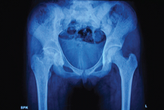
Investigations showed haemoglobin 8.7 g/dl, corrected total count 16,000/mm3, platelets3, 60,000/mm3, Erythrocyte Sedimentation Rate (ESR) 52mm (1h) and C- Reactive Protein (CRP) was 1.2mg/L. Sickling test was negative. Serum ferritin was 1000ng/ml. Frontal X-ray pelvis revealed flattening of the head of right femur with ill-defined sclerotic and lytic areas within. Right hip joint space was reduced [Table/Fig-1]. The radiographic features were suggestive of avascular necrosis of right femoral head (Steinberg class IV) [1]. Magnetic resonance imaging of hip and prothrombotic workup was not performed due to financial constraints.
Frontal radiograph of pelvis.
Paediatric orthopedic surgeon advised initial conservative management and surgery at a later date. She was treated with analgesics and crutches for non-weight bearing. At follow-up after a month her gait had improved and hip pain had considerably reduced. However, surgery was not performed as parents deferred.
Avascular necrosis of hip (Perthe’s disease) has been widely reported in the literature occurring in conjunction with homozygous sickle cell anemia and sickle thalassaemia [2]. Increased incidence of AVN of femoral head in thalassaemics when compared with general population has been reported by few authors [3–5]. The possible mechanism for this increased incidence has been attributed to a multitude of factors:- the two essential components being, anaemia and the osteoporotic bone. The chronic recurring hypoxia secondary to periodic blood transfusions augmented by ‘anaemia-associated’ hyperkinetic flow through the posterior circumflex artery could cause osteonecrosis of the femoral neck. Also, marrow hyperplasia compressing the intramedullary branches of the nutrient artery and the innately rigid and ‘less-deformable’ Red Blood Cells (RBCs) of thalassaemics could compromise the blood flow in those tiny loops of arteries. Likewise, an osteoporotic bone of a thalassaemic is prone to multiple microfractures due to the compression of the femoral neck by the acetabular roof, which serves as a nidus for the impending osteonecrosis [6]. Pre-existing inherited thrombotic disorders have also been reported in the pathogenesis of Perthe’s disease in thalassaemic patients [7]. However, work up for inherited thrombophilia and Magnetic Resonance Imaging (MRI) was not performed in the present child. A more detailed evaluation should have been carried out to look for other contributory factors. Thus, this article highlights the rarity of association of avascular necrosis of femoral head in children thalassaemia and the importance of early clinical suspicion.
[1]. Steinberg ME, Hayken GD, Steinberg DR, A quantitative system for staging avascular necrosisJ Bone Joint Surg Br 1995 77(1):34-41. [Google Scholar]
[2]. Ware HE, Brooks AP, Toye R, Berney SI, Sickle cell disease and silent avascular necrosis of the hipJ Bone Joint Surg Br 1991 73(6):947-49. [Google Scholar]
[3]. Orzincolo C, Castaldi G, Scutellari PN, Bariani L, Pinca A, Aseptic necrosis of femoral head complicating thalassaemiaSkeletal Radiol 1986 15(7):541-44. [Google Scholar]
[4]. Al-Zahrani H, Malhan H, Al-Shaalan M, Recurrent avascular necrosis of the femoral head and intramedullary bone infarcts in thalassaemia majorJ Appl Hematol 2012 3:127-28. [Google Scholar]
[5]. Madhar M, Latifi M, Aziz S, Essadki B, Fikry T, Aseptic osteonecrosis of the femoral head in thalassaemia minor: A case reportRev Chir Orthop Reparatrice Appar Mot 2005 91(2):170-72. [Google Scholar]
[6]. Katz K, Horev G, Goshen J, Tamary H, The pattern of bone disease in transfusion- dependent thalassaemia major patientsIsr J Med Sci 1994 30(8):577-80. [Google Scholar]
[7]. Levin C, Zalman L, Shalev S, Mader R, Koren A, Legg-Calve-Perthes disease, protein C deficiency, and beta-thalassaemia major: report of two casesJ Pediatr Orthop 2000 20(1):129-31. [Google Scholar]